. 2004 Apr;2(4):E90.
doi: 10.1371/journal.pbio.0020090. Epub 2004 Apr 13.

Mimotopes for alloreactive and conventional T cells in a peptide-MHC display library

Affiliations

Mimotopes for alloreactive and conventional T cells in a peptide-MHC display library

Frances Crawford et al. PLoS Biol. 2004 Apr.

Abstract

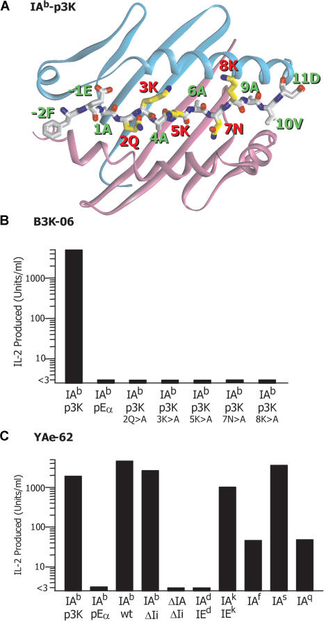
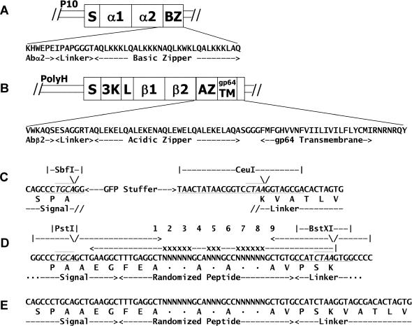
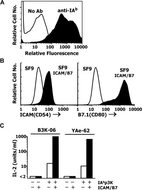
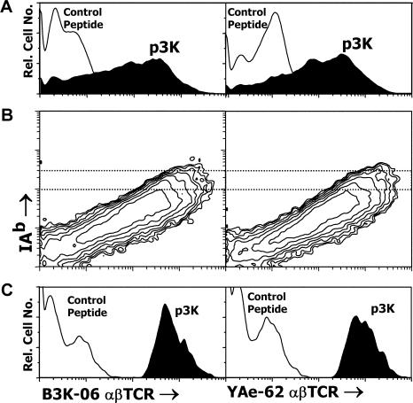
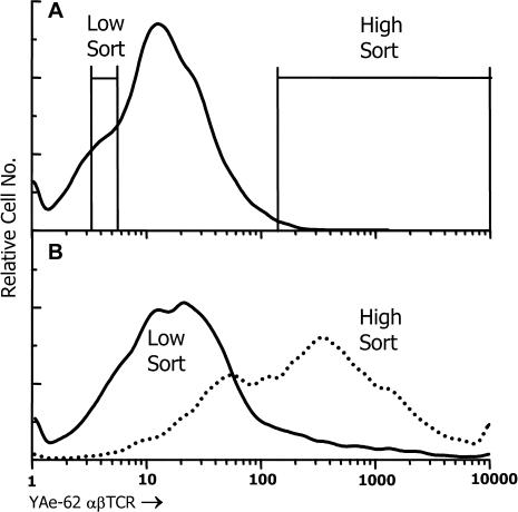
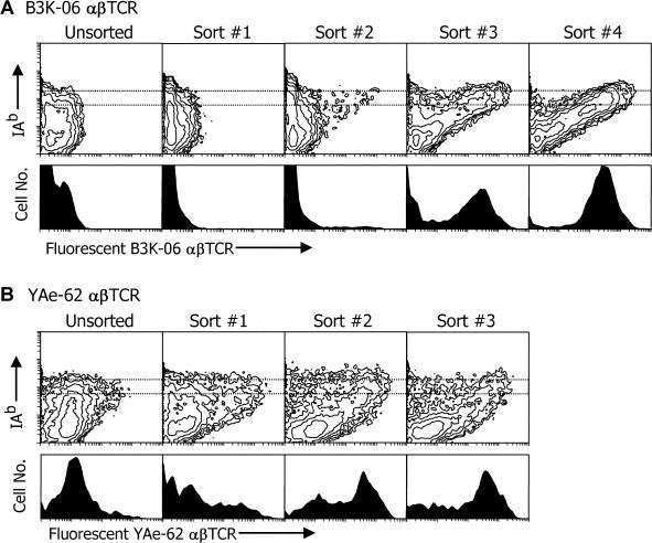
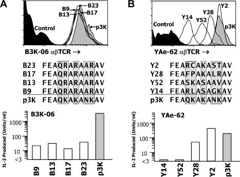
The use of peptide libraries for the identification and characterization of T cell antigen peptide epitopes and mimotopes has been hampered by the need to form complexes between the peptides and an appropriate MHC molecule in order to construct a complete T cell ligand. We have developed a baculovirus-based peptide library method in which the sequence encoding the peptide is embedded within the genes for the MHC molecule in the viral DNA, such that insect cells infected with virus encoding a library of different peptides each displays a unique peptide-MHC complex on its surface. We have fished in such a library with two different fluorescent soluble T cell receptors (TCRs), one highly peptide specific and the other broadly allo-MHC specific and hypothesized to be much less focused on the peptide portion of the ligand. A single peptide sequence was selected by the former alphabetaTCR that, not unexpectedly, was highly related to the immunizing peptide. As hypothesized, the other alphabetaTCR selected a large family of peptides, related only by a similarity to the immunizing peptide at the p5 position. These findings have implications for the relative importance of peptide and MHC in TCR ligand recognition. This display method has broad applications in T cell epitope identification and manipulation and should be useful in general in studying interactions between complex proteins.


Conflict of interest statement

The authors have declared that no conflicts of interest exist.

Figures

Figure 1
Figure 1. Structure of IAb-p3K and Properties of T Cell Hybridomas Reactive to It
(A) Ribbon structure of the α1 and β1 domains of IAb with a wire-frame representation of the bound p3K peptide (Liu et al. 2002). Amino acids labeled in red are the five central peptide amino acids available for αβTCR interaction. (B) The figure shows the response of 105 B3K-06 hybridoma cells to various peptides presented by 105 IAb-bearing APCs, LB-15.13. (C) The figure shows the response of the T cell hybridoma YAe-62 to various MHCII molecules. In each case, 105 hybridoma cells were incubated overnight with MHCII presented in various ways. For IAb-p3K, soluble IAb-p3K was immobilized in the culture well before the addition of the hybridoma cells. In other cases, 106 spleen cells were used directly as APCs without additional peptide antigen. For pEα, the spleen cells came from IAb-pEα/ΔIAβ/ΔIi mice (Ignatowicz et al. 1996). For wild-type IAb and allo-MHCII, the spleen cells came from H-2 congenic mice on the C57BL/10 background. Finally, spleen cells from ΔIAβ/ΔIi C57BL/6 mice were used.
Figure 2
Figure 2. Constructions Used in These Experiments
(A and B) Previously described constructions (Rees et al. 1999) for the coexpression in a single baculovirus of soluble version of the α (A) and β (B) chains of IAb were modified as described in the Materials and Methods to anchor the molecule on the surface of infected insect cells. (C) The construction was further modified as described in the Materials and Methods to disrupt the IAb β chain with sequence encoding enhanced GFP flanked by sites for the enzymes SbfI and CeuI. (D and E) A degenerate DNA fragment was produced by PCR (D) and cloned into the construct replacing the GFP-encoding sequence (E) as described in the Materials and Methods.
Figure 3
Figure 3. Functional Display of IAb-p3K on the Surface of Insect Cells
(A) Sf9 insect cells were infected with baculovirus encoding a membrane-bound form of IAb-p3K. After 3 d, the surface expression of IAb-p3K was detected with an anti-IAb mAb using flow cytometry. (B) The genes for mouse ICAM (CD54) and B7.1 (CD80) were cloned into an insect cell expression plasmid as described in the Materials and Methods. The plasmids were used to cotransfect Sf9 cells, and a stable transfectant (Sf9-ICAM/B7.1) was cloned expressing both proteins detected with mAbs using flow cytometry. (C) Either Sf9 (open bars) or Sf9-ICAM/B7.1 (closed bars) cells were infected with baculovirus expressing IAb-p3K. After 3 d, the infected insect cells were used as APCs to stimulate IL-2 production from B3K-06 and YAe-62. Uninfected cells were used as negative controls.
Figure 4
Figure 4. Detection of IAb-p3K-Expressing Insect Cells with Polyvalent, Fluorescent αβTCRs
(A) Sf9 insect cells were infected with baculovirus encoding IAb bound either to p3K (filled histogram) or a control peptide (FEAPVAAALHAV) (unfilled histogram). After 3 d, the infected insect cells were incubated with polyvalent, fluorescent soluble αβTCRs from B3K-06 or YAe-62. The binding of each αβTCR was assessed by flow cytometry. (B) Cells, prepared as in (A), were simultaneously analyzed with fluorescent αβTCRs and a mAb specific for IAb (17–227) that does not interfere with αβTCR–IAb interaction. (C) The binding of the αβTCRs is shown only for those infected insect cells that bear a high level of surface IAb (dotted region in [B]).
Figure 5
Figure 5. Recovery of IAb-p3K Virus-Infected Cells with Fluorescent αβTCR
(A) Sf9 cells were infected with a mixture of virus, 99% of which encoded a control protein (a TCR β chain linked to the gp64 transmembrane/cytoplasmic tail) and 1% of which encoded IAb-p3K. After 3 d, the infected cells were analyzed as in Figure 3A for binding fluorescent αβTCR from YAe-62. The 1% of the infected cells with the brightest fluorescence was sorted (high sort, 15,700 cells). As a control, a similar number of cells that fluoresced as dully as the background fluorescence were also sorted (low sort). (B) The sorted cells were incubated with fresh Sf9 insect cells to allow propagation of the viruses and production of new stocks. The stocks were used to infect new Sf9 cells, and after 3 d the analysis of αβTCR binding was repeated.
Figure 6
Figure 6. Summary of Successive Screening of the IAb–Peptide Libraries with Fluorescent αβTCRs
Sf9 insect cells (1 × 10⁷ to 1.5 × 10⁷) were infected at a MOI of approximately 1 with an aliquot of baculovirus encoding the IAb–peptide library. After 3 d, the infected cells were analyzed for binding the αβTCR of either B3K-06 or YAe-62. Either obviously fluorescent cells or the brightest 1% of the cells were sorted (2 × 10⁴ to 8 × 10⁴ cells) and added to 3 × 10⁶ fresh Sf9 cells to propagate and reexpress the viruses contained in the sorted cells. These infected cells were then reanalyzed and sorted using the fluorescent αβTCRs. This process was repeated until no further enrichment of αβTCR binding was seen. In most cases, the reanalysis was done directly from the cells that were cocultured with the sorted cells. In a few cases, an intermediate viral stock was made and then used to infect additional Sf9 cells. The turn around time per cycle was 4–7 d. The figure shows the reanalysis in a single experiment of the initial viral stocks and all of the various intermediate enriched viral stocks. Sf9 cells were infected at an MOI of less than 1 with the viral stocks and analyzed as in Figure 4 for either B3K-06 (A) or YAe-62 (B) αβTCR binding.
Figure 7
Figure 7. Analysis of Baculovirus Clones from the αβTCR-Enriched IAb–Peptide Library
(A) Sf9 cells were infected with stock from four baculovirus clones (B9, B13, B17, and B23) isolated from the virus pool enriched with the αβTCR of B3K-06. After 3 d, an aliquot of cells from each infection was analyzed as in Figure 4 to assure uniform binding of the fluorescent B3K-06 αβTCR (top). Viral DNAs prepared from other aliquots of the cells were used as templates in a PCR with oligonucleotides that flanked the DNA encoding the IAb-bound peptide. The fragment was sequenced directly with a third internal oligonucleotide (middle). The clone stock was then used to infect Sf9-ICAM/B7.1 cells. After 3 d, the infected cells were used as APCs for B3K-06 production of IL-2 (bottom). Virus encoding IAb-p3K was used as a positive control. Virus encoding pEα was used as the negative control. (B) Same as (A), but using YAe-62 and clones (Y2, Y14, Y28, Y52) derived from the IAb–peptide library using the YAe-62 αβTCR.

Similar articles

Cited by

References

    1. Boder ET, Wittrup KD. Yeast surface display for directed evolution of protein expression, affinity, and stability. Methods Enzymol. 2000;328:430–444. - PubMed
    1. Brock R, Wiesmuller KH, Jung G, Walden P. Molecular basis for the recognition of two structurally different major histocompatibility complex/peptide complexes by a single T-cell receptor. Proc Natl Acad Sci U S A. 1996;93:13108–13113. - PMC - PubMed
    1. Brophy SE, Holler PD, Kranz DM. A yeast display system for engineering functional peptide–MHC complexes. J Immunol Methods. 2003;272:235–246. - PubMed
    1. Cai Z, Brunmark A, Jackson MR, Loh D, Peterson PA, et al. Transfected Drosophila cells as a probe for defining the minimal requirements for stimulating unprimed CD8+ T cells. Proc Natl Acad Sci U S A. 1996;93:14736–14741. - PMC - PubMed
    1. Crawford F, Kozono H, White J, Marrack P, Kappler J. Detection of antigen-specific T cells with multivalent soluble class II MHC covalent peptide complexes. Immunity. 1998;8:675–682. - PubMed

Publication types

MeSH terms