. 2004 May;112(1):54-63.
doi: 10.1111/j.1365-2567.2004.01857.x.

Major histocompatibility complex class I-restricted alloreactive CD4+ T cells

Affiliations

Major histocompatibility complex class I-restricted alloreactive CD4+ T cells

Louise H Boyle et al. Immunology. 2004 May.

Abstract

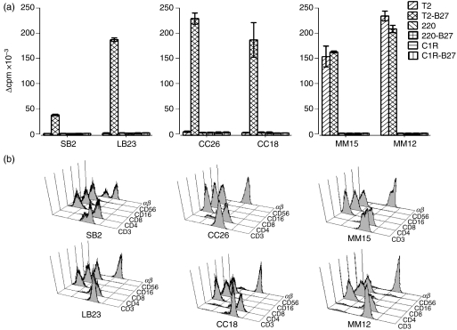
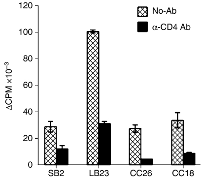
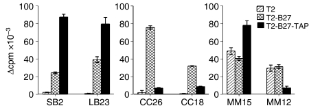
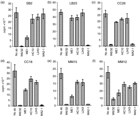
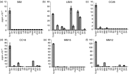
Although it is well established that CD4+ T cells generally recognize major histocompatibility complex (MHC) class II molecules, MHC class I-reactive CD4+ T cells have occasionally been reported. Here we describe the isolation and characterization of six MHC class I-reactive CD4+ T-cell lines, obtained by co-culture of CD4+ peripheral blood T cells with the MHC class II-negative, transporter associated with antigen processing (TAP)-negative cell line, T2, transfected with human leucocyte antigen (HLA)-B27. Responses were inhibited by the MHC class I-specific monoclonal antibody (mAb), W6/32, demonstrating the direct recognition of MHC class I molecules. In four cases, the restriction element was positively identified as HLA-A2, as responses by these clones were completely inhibited by MA2.1, an HLA-A2-specific mAb. Interestingly, three of the CD4+ T-cell lines only responded to cells expressing HLA-B27, irrespective of their restricting allele, implicating HLA-B27 as a possible source of peptides presented by the stimulatory MHC class I alleles. In addition, these CD4+ MHC class I alloreactive T-cell lines could recognize TAP-deficient cells and therefore may have particular clinical relevance to situations where the expression of TAP molecules is decreased, such as viral infection and transformation of cells.


Figures

Figure 1
Figure 1
CD4+ T-cell lines proliferate in response to T2-B27 (a T2-cell line transfected with HLA-B*2705). (a) Proliferation is expressed as Δ counts per minute (c.p.m.) of CD4+ T-cell lines to a panel of B27+ and B27 stimulator cell lines. Data shown are the means of duplicate wells ± standard deviation (SD). (b) Flow cytometric analysis of the phenotype of the six T2-B27-reactive T-cell lines.
Figure 2
Figure 2
Responses to T2-B27 (a T2-cell line transfected with HLA-B*2705) are CD4 dependent. Proliferative responses are displayed as Δ counts per minute (c.p.m.) to T2-B27 in the presence or absence of a blocking monoclonal antibody (mAb) specific for CD4. Data shown are the means of triplicate results ± standard deviation (SD).
Figure 3
Figure 3
Differential responses to T2-B27 (a T2-cell line transfected with HLA-B*2705) on reconstitution of TAP molecules. Proliferative responses, displayed as Δ counts per minute (c.p.m.) of the CD4+ T-cell lines to T2, T2-B27 and T2-B27-TAP (T2-B27 reconstituted with TAP1 and TAP2). Data shown represent the mean values of triplicate wells ± standard deviation (SD).
Figure 4
Figure 4
Inhibition of responses to T2-B27 (a T2-cell line transfected with HLA-B*2705) by major histocompatibility complex (MHC) class I-specific monoclonal antibodies (mAbs). Proliferative responses, displayed as Δ counts per minute (c.p.m.), to T2-B27 in the presence or absence of 10 µg/ml of the MHC class I-specific mAb, W6/32, the human leucocyte antigen (HLA)-B27-specific mAb, ME1, the free heavy chain-specific mAb, HC10, the HLA-DR-specific mAb, L243, and the HLA-A2-specific mAb, MA2.1. Data shown are the means of triplicate wells ± standard deviation (SD).
Figure 5
Figure 5
Proliferative responses are observed to the Epstein–Barr virus-transformed lymphoblastoid cell line (EBV-LCL) by three of the major histocompatibility complex (MHC) class I-reactive CD4+ T-cell lines. Proliferative responses are expressed as Δ counts per minute (c.p.m.) to a panel of EBV-LCL. Proliferative responses to T2-B27 (a T2-cell line transfected with HLA-B*2705) are shown as a control. Data shown represent the mean values of triplicate results.

References

    1. Spits H, Ijssel H, Thompson A, de Vries JE. Human T4+ and T8+ cytotoxic T lymphocyte clones directed at products of different class II major histocompatibility complex loci. J Immunol. 1983;131:678–83. - PubMed
    1. Kirberg J, Baron A, Jakob S, Rolink A, Karjalainen K, von Boehmer H. Thymic selection of CD8+ single positive cells with a class II major histocompatibility complex-restricted receptor. J Exp Med. 1994;180:25–34. - PMC - PubMed
    1. Suzuki H, Eshima K, Takagaki Y, et al. Origin of a T cell clone with a mismatched combination of MHC restriction and coreceptor expression. J Immunol. 1994;153:4496–507. - PubMed
    1. Heemskerk MH, Schilham MW, Schoemaker HM, Spierenburg G, Spaan WJ, Boog CJ. Activation of virus-specific major histocompatibility complex class II-restricted CD8+ cytotoxic T cells in CD4-deficient mice. Eur J Immunol. 1995;25:1109–12. - PMC - PubMed
    1. Bot A, Casares S, Bot S, von Boehmer H, Bona C. Cellular mechanisms involved in protection against influenza virus infection in transgenic mice expressing a TCR receptor specific for class II hemagglutinin peptide in CD4+ and CD8+ T cells. J Immunol. 1998;160:4500–7. - PubMed

Publication types

MeSH terms

Substances