

Counteraction of urea-induced protein denaturation by trimethylamine N-oxide: a chemical chaperone at atomic resolution
- PMID: 15096583
- PMCID: PMC404062
- DOI: 10.1073/pnas.0308633101
Counteraction of urea-induced protein denaturation by trimethylamine N-oxide: a chemical chaperone at atomic resolution
Abstract
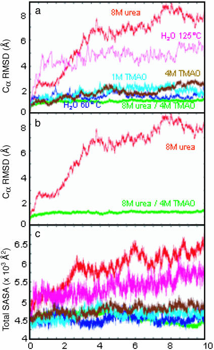
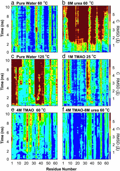
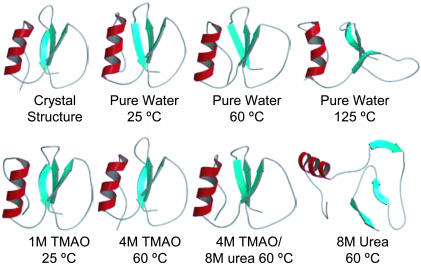
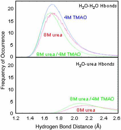
Proteins are very sensitive to their solvent environments. Urea is a common chemical denaturant of proteins, yet some animals contain high concentrations of urea. These animals have evolved an interesting mechanism to counteract the effects of urea by using trimethylamine N-oxide (TMAO). The molecular basis for the ability of TMAO to act as a chemical chaperone remains unknown. Here, we describe molecular dynamics simulations of a small globular protein, chymotrypsin inhibitor 2, in 8 M urea and 4 M TMAO/8 M urea solutions, in addition to other control simulations, to investigate this effect at the atomic level. In 8 M urea, the protein unfolds, and urea acts in both a direct and indirect manner to achieve this effect. In contrast, introduction of 4 M TMAO counteracts the effect of urea and the protein remains well structured. TMAO makes few direct interactions with the protein. Instead, it prevents unfolding of the protein by structuring the solvent. In particular, TMAO orders the solvent and discourages it from competing with intraprotein H bonds and breaking up the hydrophobic core of the protein.
Figures
References
-
- Yancey, P. H., Clark, M. E., Hand, S. C., Bowlus, R. D. & Somero, G. N. (1982) Science 217, 1214-1222. - PubMed
-
- Withers, P. C., Morrison, G. & Guppy, M. (1994) Physiol. Zool. 67, 693-705.
-
- Barton, K. N., Buhr, M. M. & Ballantyne, J. S. (1999) Am. J. Physiol. 276, R397-R406. - PubMed
-
- Yancey, P. H., Blake, W. R. & Conley, J. (2002) Comp. Biochem. Physiol. A Mol. Integr. Physiol. 133, 667-676. - PubMed
-
- Yancey, P. H., Fyfe-Johnson, A. L., Kelly, R. H., Walker, V. P. & Aunon, M. T. (2001) J. Exp. Zool. 289, 172-176. - PubMed
Publication types
MeSH terms
Substances
Grants and funding
LinkOut - more resources
Full Text Sources
Other Literature Sources
