. 2004 May 18;101(20):7530-5.
doi: 10.1073/pnas.0401634101. Epub 2004 Apr 19.

An aminoacyl-tRNA synthetase-like protein encoded by the Escherichia coli yadB gene glutamylates specifically tRNAAsp

Affiliations

An aminoacyl-tRNA synthetase-like protein encoded by the Escherichia coli yadB gene glutamylates specifically tRNAAsp

Daniel Y Dubois et al. Proc Natl Acad Sci U S A. .

Abstract

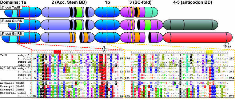
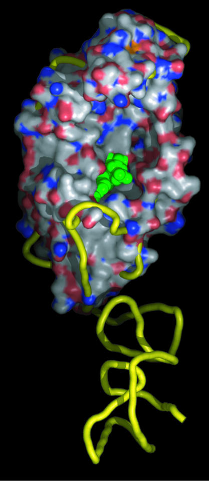
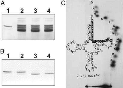
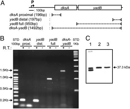
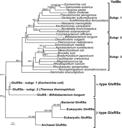
The product of the Escherichia coli yadB gene is homologous to the N-terminal part of bacterial glutamyl-tRNA synthetases (GluRSs), including the Rossmann fold with the acceptor-binding domain and the stem-contact fold. This GluRS-like protein, which lacks the anticodon-binding domain, does not use tRNA(Glu) as substrate in vitro nor in vivo, but aminoacylates tRNA(Asp) with glutamate. The yadB gene is expressed in wild-type E. coli as an operon with the dksA gene, which encodes a protein involved in the general stress response by means of its action at the translational level. The fate of the glutamylated tRNA(Asp) is not known, but its incapacity to bind elongation factor Tu suggests that it is not involved in ribosomal protein synthesis. Genes homologous to yadB are present only in bacteria, mostly in Proteobacteria. Sequence alignments and phylogenetic analyses show that the YadB proteins form a distinct monophyletic group related to the bacterial and organellar GluRSs (alpha-type GlxRSs superfamily) with ubiquitous function as suggested by the similar functional properties of the YadB homologue from Neisseria meningitidis.


Figures

Fig. 1.
Fig. 1.
Comparison of the sequences of YadB proteins with GluRSs and GlnRSs showing the modular arrangements and the structural relationships of the three families of proteins. The catalytic domain (1a and 1b), the acceptor stem-binding domain [2 (Acc. Stem BD)], and the stem-contact fold domain [3 (SC-fold)] are blue, pastel blue, and purple, respectively. The anticodon-binding domain(s) of GluRSs and GlnRSs are gray (α-type, 4–5) and light red (β-type, 4), respectively. Insertions specificto α- and β-type GlxRSs are light green and orange, respectively. The YadB-specific C-terminal sequence is turquoise. The red and yellow strips above the sequences indicate the location of the class I aaRS sequence motifs “HIGH” and “KMSK”, respectively. A structure-based alignment of amino acid consensus sequences of the regions containing these two motifs are presented in red and yellow boxes. The consensus sequences represent the three YadB subgroups, subgroups 1 and 2 of bacterial and organellar (B/O) α-type GluRSs, and the β-type GlxRSs (archaeal and eukaryal GluRSs, and bacterial and eukaryal GlnRSs). Aligned individual sequences of E. coli YadB, GluRS and GlnRS and T. thermophilus GluRS are also presented. Residues are presented by colored characters: highlighted for 100% conservation, bold for 75–99%, normal for 50–74%, and a dot (or lowercase character in the individual sequences) for <50% conservation. The black arrow points at a conserved residue distinct in YadBs and GlxRSs (see the text).
Fig. 2.
Fig. 2.
Three-dimensional structure of E. coli YadB and comparison with homologous GluRS. The structures of YadB has been superimposed on that of the T. thermophilus GluRS (1GLN, 6). The figure displays YadB surface (C, white; O, red; N, blue) and GluRS ribbon trace (yellow). The modeled aminoacyl-adenylate is emphasized in green. The coordinates of YadB are deposited in the Protein Data Bank (www.pdb.org; PDB ID code 1NZJ). Figure 2 was made with pymol.
Fig. 3.
Fig. 3.
Identification and characterization of the tRNA charged by YadB. (A) PAGE in 40 mM Mops (pH 6.5)/10 mM sodium acetate/1 mM EDTA. Lane 1, 2 μg of purified RNA charged with l-[14C]Glu by E. coli YadB; lanes 2–4, 40 μgof unfractionated E. coli tRNA charged with l-[14C]Glu by E. coli YadB or GluRS or with l-[14C]Gln by GlnRS, respectively. The RNAs were stained with methylene blue. (B) PhosphorImager exposure of the gel shown in A. (C) Homochromatogram of RNA fragment specific of E. coli tRNAAsp and cloverleaf fold of this tRNA. The oligonucleotide identifying tRNAAsp is displayed on a black background.
Fig. 4.
Fig. 4.
In vivo expression of E. coli YadB gene. (A) Physical map of the E. coli dksA-yadB region showing the localization of the RT-PCR amplification targets. The promoter for dksA identified by Kang and Craig (47) is indicated by the letter P. (B) Expression of the YadB mRNA in E. coli K12 grown in LB medium/agarose gel electrophoresis of the products of the RT-PCR reactions and of controls without the reverse polymerase (R.T.). (C) Expression of the YadB protein. Lanes 1–3, analysis by Western blot of, respectively, 10 ng of overexpressed pure YadB and 9 μg of crude and S100 extracts from wild-type E. coli.
Fig. 5.
Fig. 5.
Rooted phylogenetic tree of YadB proteins and GluRS/GlnRS representatives. Bootstrap values (>50%) calculated from 400 replicates in the maximum-parsimony analysis, 1,000 replicates in the neighbor-joining analysis, and 5,000 puzzling steps in the maximum-likelihood analysis are indicated at their corresponding nodes separated by ′/′ in that specific order. The tree is rooted with P. horikoshi class I LysRS (not shown).

Comment in

References

    1. Woese, C. R., Olsen, G. J., Ibba, M. & Söll, D. (2000) Microbiol. Mol. Biol. Rev. 64, 202-236. - PMC - PubMed
    1. Schimmel, P. & Ribas De Pouplana, L. (2000) Trends Biochem. Sci. 25, 207-209. - PubMed
    1. Francklyn, C. S., Perona, J. J., Pütz, J. & Hou, Y. M. (2002) RNA 8, 1363-1372. - PMC - PubMed
    1. Fujita, N., Mori, H., Yura, T. & Ishihama, A. (1994) Nucleic Acids Res. 22, 1637-1639. - PMC - PubMed
    1. Blattner, F. R., Plunkett, G., III, Bloch, C. A., Perna, N. T., Burland, V., Riley, M., Collado-Vides, J., Glasner, J. D., Rode, C. K., Mayhew, G. F., et al. (1997) Science 277, 1453-1474. - PubMed

Publication types

MeSH terms

LinkOut - more resources