. 2004 Apr 27;101(17):6502-7.
doi: 10.1073/pnas.0401711101. Epub 2004 Apr 19.

Essential role of Sox9 in the pathway that controls formation of cardiac valves and septa

Affiliations

Essential role of Sox9 in the pathway that controls formation of cardiac valves and septa

Haruhiko Akiyama et al. Proc Natl Acad Sci U S A. .

Abstract

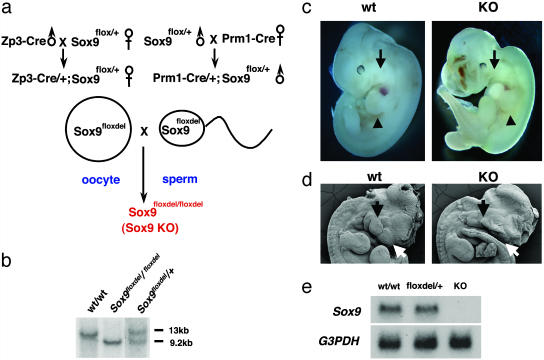
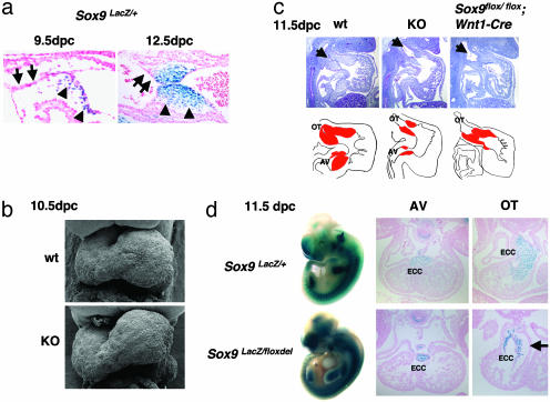
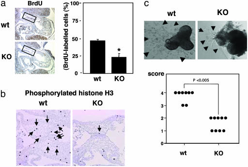
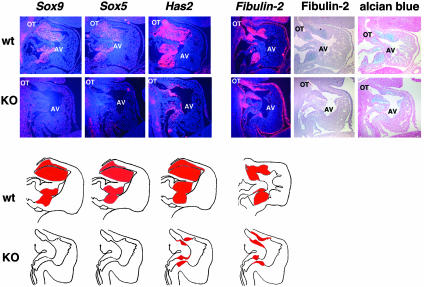
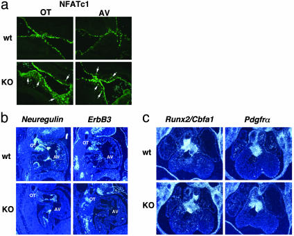
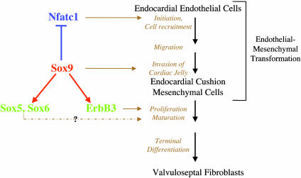
Epithelial-mesenchymal transformation is a critical developmental process reiterated in multiple organs throughout embryogenesis. Formation of endocardial cushions, primordia of valves and septa, is a classic example of epithelial-mesenchymal transformation. Several gene mutations are known to affect cardiac valve formation. Sox9 is activated when endocardial endothelial cells undergo mesenchymal transformation and migrate into an extracellular matrix, called cardiac jelly, to form endocardial cushions. In Sox9-null mutants, endocardial cushions are markedly hypoplastic. In these mutants, Nfatc1 is ectopically expressed and no longer restricted to endothelial cells. Further, Sox9-deficient endocardial mesenchymal cells fail to express ErbB3, which is required for endocardial cushion cell differentiation and proliferation. Our results reveal a succession of molecular steps in the pathway of endocardial cushion development. We propose that loss of Sox9 inhibits epithelial-mesenchymal transformation after delamination and initial migration, but before definitive mesenchymal transformation.


Figures

Fig. 1.
Fig. 1.
Targeting strategy for Sox9-null embryos. (a) The mating scheme used to generate Sox9-null embryos. (b) Southern blot analysis of fetal genomic DNA. Genomic DNA isolated from the yolk sac was digested with BamHI and then hybridized with the 3′ probe. The wild-type and floxdel alleles were detected as 13-kb and 9.2-kb fragments, respectively. (c) The gross appearance of wild-type (wt) and Sox9-null (KO) embryos at 11.5 dpc. The arrows indicate the first branchial arch. The arrowheads indicate the forelimb buds. (d) Morphological analysis of craniofacial regions of wild-type and Sox9-null embryos at 11.5 dpc by using a scanning electron microscope. The white and black arrows indicate frontonasal mass and branchial arches, respectively. (e) Northern blot analysis of Sox9 mRNA expression in wild-type and Sox9-null embryos at 11.5 dpc.
Fig. 2.
Fig. 2.
Hypoplastic endocardial cushions in Sox9-null embryos. (a) Sox9 expression in endocardial cushions of Sox9 heterozygous embryos at 9.5 and 12.5 dpc. The arrows and the arrowheads indicate endothelial cells and mesenchymal cells, respectively. (b) Morphological analysis of the heart of wild-type and Sox9-null embryos at 10.5 dpc by using scanning electron microscopy. (c) Hematoxylin and Treosin staining of Sox9-null embryos and the Sox9 conditional null mutant embryos at 11.5 dpc. The arrows indicate the outflow tract of the heart. In graphical drawings, red denotes endocardial cushions. Five independent experiments were performed and gave similar results. (d) Distribution of Sox9-null cells. Whole-mount X-Gal staining of Sox9 heterozygous (Sox9lacZ/+) and Sox9-null (Sox9lacZ/floxdel) embryos at 11.5 dpc, and transverse sections of Sox9 heterozygous (Sox9lacZ/+) and Sox9-null (Sox9lacZ/floxdel) embryos at 11.5 dpc stained with X-Gal. The arrow indicates the acellular cardiac jelly in Sox9-null embryos. ECC, endocardial cushion; OT, outflow tract. Three independent experiments were performed and gave similar results.
Fig. 3.
Fig. 3.
Cell proliferation and morphogenesis of AV canal endocardial cushion cells. (a) BrdUrd incorporation was reduced in endocardial cushion cells (boxed regions) in Sox9-null embryos. Statistical significance was assessed by one-way analysis of variance and unpaired Student's t test. Values shown are the mean + SD of four embryos. *, Statistically significant difference between wild-type and Sox9-null embryos at P < 0.001. (b) Immunohistochemistry of phosphorylated histone H3 revealed marked reduction of the number of phosphorylated histone H3-positive cells in endocardial cushion of Sox9-null embryos. These results were confirmed with two wild-type (wt) and two Sox9-null mutant (KO) embryos. (c) AV canal explant cultures showed markedly reduced transformation of endothelial cells into mesenchyme. The arrowheads indicate migrating endothelial cells and transformed mesenchymal cells. Eight wild-type and nine Sox9-null mutant explant cultures were examined. Statistical analysis used the Wilcoxon rank sum test (17).
Fig. 4.
Fig. 4.
Histological analysis in endocardial cushions of Sox9-null embryos. Hearts of wild-type (wt) and Sox9-null (KO) embryos at 11.5 dpc were hybridized with probes for Sox9, Sox5, Has2, and Fibulin-2. In graphical drawings, red denotes the expression domains of these genes in endocardial cushions. The expression of Sox5 was abolished. Alcian blue staining and immunohistochemistry of fibulin-2 showed marked reduction of matrix accumulation in endocardial cushion of Sox9-null embryos. OT, outflow tract. Three independent experiments were performed and gave similar results.
Fig. 5.
Fig. 5.
Histological analysis in endocardial cushions of Sox9-null embryos. (a) Immunofluorescence of Nfatc1 in sections of wild-type (wt) and Sox9-null (KO) embryos at 11.5 dpc. The arrows indicate expression of Nfatc1 in mesenchymal cells of endocardial cushions in Sox9-null mutants. (b and c) Hearts of wild-type and Sox9-null embryos at 11.5 dpc were hybridized with probes for Neuregulin, ErbB3, Runx2/Cbfa1, and Pdgfrα. The expression of ErbB3 was abolished. OT, outflow tract. Three independent experiments were performed and gave similar results.
Fig. 6.
Fig. 6.
Model of the functions of Sox9 in the differentiation of endocardial cushion cells. Sox9 is expressed in mesenchymal cells, not in endothelial cells of endocardial cushions, prevents expression in mesenchymal cells of Nfatc1, which is expressed in endocardial endothelial cells, and is needed at an early step for endocardial cushion formation. Sox9 is required for expression of ErbB3, which is needed for proliferation and differentiation of endocardial cushion cells. Sox9 also activates Sox5 and Sox6.

References

    1. Zhao, Q., Eberspaecher, H., Lefebvre, V. & De Crombrugghe, B. (1997) Dev. Dyn. 209, 377-386. - PubMed
    1. Ng, L. J., Wheatley, S., Muscat, G. E., Conway-Campbell, J., Bowles, J., Wright, E., Bell, D. M., Tam, P. P., Cheah, K. S. & Koopman, P. (1997) Dev. Biol. 183, 108-121. - PubMed
    1. Wagner, T., Wirth, J., Meyer, J., Zabel, B., Held, M., Zimmer, J., Pasantes, J., Bricarelli, F. D., Keutel, J., Hustert, E., et al. (1994) Cell 79, 1111-1120. - PubMed
    1. Foster, J. W., Dominguez-Steglich, M. A., Guioli, S., Kowk, G., Weller, P. A., Stevanovic, M., Weissenbach, J., Mansour, S., Young, I. D., Goodfellow, P. N., et al. (1994) Nature 372, 525-530. - PubMed
    1. Houston, C. S., Opitz, J. M., Spranger, J. W., Macpherson, R. I., Reed, M. H., Gilbert, E. F., Herrmann, J. & Schinzel, A. (1983) Am. J. Med. Genet. 15, 3-28. - PubMed

Publication types