. 2004 Apr 27;101(17):6373-8.
doi: 10.1073/pnas.0401855101. Epub 2004 Apr 19.

Gonadotropin-regulated testicular RNA helicase (GRTH/Ddx25) is essential for spermatid development and completion of spermatogenesis

Affiliations

Gonadotropin-regulated testicular RNA helicase (GRTH/Ddx25) is essential for spermatid development and completion of spermatogenesis

Chon-Hwa Tsai-Morris et al. Proc Natl Acad Sci U S A. .

Abstract

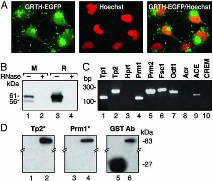
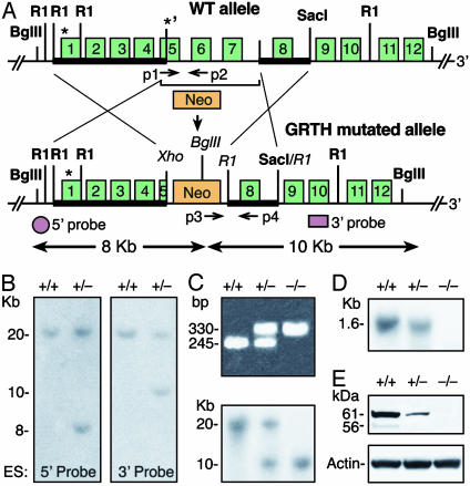
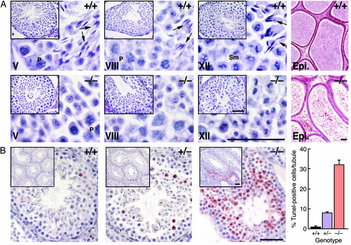
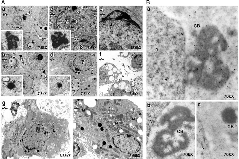
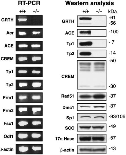
Gonadotropin-regulated testicular RNA helicase (GRTH/Ddx25), a member of the DEAD-box protein family, is a testis-specific gonadotropin-regulated RNA helicase that is present in Leydig cells and germ cells (meiotic spermatocytes and spermatids). In this study, we observed that GRTH is present in the nucleus, cytoplasm and chromatoid body of germ cells, and is an integral component of messenger ribonuclear protein particles. Male mice with a null mutation in the GRTH gene displayed normal gonadotropin and androgen profiles. However, they were sterile, with azoospermia caused by a complete arrest of spermiogenesis at step 8 of round spermatids and failure to elongate. Round spermatids of the null mice showed marked diminution in the size of chromatoid bodies. The transcription of relevant messages was not altered, but their translation was abrogated in a selective manner. Protein expression of transition proteins 1 and 2 and angiotensin-converting enzyme was completely absent, whereas that of the transcriptional activator cAMP responsive element modulator was intact. These findings indicate that GRTH participates in translational-associated events during germ cell development. Although significant apoptosis was present at the metaphase of meiosis in the GRTH-null mice, spermatogenesis proceeded to step 8 of spermiogenesis when complete arrest occurred. This progression may relate to compensatory gene function(s) and/or the observed up-regulation of DNA repair proteins Rad51 and Dmc1. This study (i) demonstrates that GRTH is essential for completion of spermatogenesis, (ii) provides insights into intrinsic requirements for spermiogenesis, and (iii) establishes a model for studies of male infertility and contraception.


Figures

Fig. 1.
Fig. 1.
Cellular localization of GRTH protein and its association with mRNP. (A) Confocal analysis of overexpressed GRTH–GFP fusion protein in COS-1 cells (Left), nuclear staining by Hoechst 33342 (Middle), and merge image (Right). (B) Western analysis of oligo(dT)-purified mRNA–protein complex from mouse (M) and rat (R) testis. (C) RT-PCR analysis of immunoprecipitated testicular GRTH complexes with germ cell-specific mRNAs and hypoxanthine guanine phosphoribosyl transferase (Hprt; ubiquitously expressed). Fsc1, fibrous sheath component-1. Odf1, outer dense fiber. Acr, acrosin. (D) Northwestern analysis (Left) of GRTH–GST fusion protein binding to in vitro transcribed 3′UTR 32P-labeled RNA probe (*) of Tp2 (lane 2) and Prm1 (lane 4). GST, negative control (lanes 1 and 3). Western blot (Right) with a GST antibody shows the presence of GRTH–GST (83 kDa) and GST (27 kDa).
Fig. 2.
Fig. 2.
Targeted disruption of the GRTH gene. (A) Targeted gene construction. (Upper) Wild-type mouse GRTH gene allele. *, ATGs. Bold line indicates the homologous 5′ arm (exons 1–4 and part of exon 5) and the 3′ arm (2.9-kb fragment with exon 8, 5′ and 3′ intron sequences) were subcloned at NotI/Xho and R1 site, respectively, in the targeting vector pPNT with (Neo) and (TK) selection markers. (Lower) The GRTH mutated homologus recombinant allele. Shown are the 5′ and 3′ probes used for Southern analysis and the PCR primers used for genotype screening (p1-p4). (B) Screening ES clones by Southern analysis. Positive homologous recombinant clones were identified with BglII digestion (predicted size: wild-type, 20 kb; recombinant allele with 5′ probe, 8 kb, and 3′ probe, 10 kb). (C) Genotyping of offspring mice by PCR (Upper). Predicted size: GRTH+/+, 245 bp (p1/p2); GRTH+/-, 245 bp (p1/p2) and 330 bp (p3/p4); and GRTH-/- mice, 330 bp (p3/p4). Positive clones were confirmed by Southern analysis by using a 3′ probe (Lower). (D and E) Northern (D) and Western (E) analyses of testis from adult GRTH+/+, GRTH+/-, and GRTH-/- mice.
Fig. 3.
Fig. 3.
Complete arrest of spermatogenesis in GRTH-/- mice. (A) Periodic acid/Schiff reagent-stained sections from 16-week-old GRTH+/+ and GRTH -/- mice testis. (Scale bar, 0.5 μm.) Elongated spermatids and mature sperm were completely absent in the testis of GRTH-/- animals, and no mature sperm was observed in the epididymis. Arrows indicate elongated spermatids. P, pachytene spermatocytes. Sm, spermatocytes in the metaphase of meiosis. (B) TUNEL assay. Error bars indicate mean ± SEM positive cells per tubule. (Scale bar, 0.5 μm.)
Fig. 4.
Fig. 4.
EM analysis of GRTH+/+ and GRTH-/- mouse testis. (A) Spermatids of GRTH+/+ and GRTH-/- at step 2/3 (a and b) and at step 7/8 (c and d) of spermiogenesis. White arrows indicate round spermatid CB. (Inset) CB at higher magnification (×40,000). *, Acrosomal vesicles noted in step 2/3. **, Typical acrosomal structure noted in step 7/8. (e) Black arrow indicates abnormal acrosome in GRTH -/- mice after arrest. (f) Degenerating round spermatids at stage XII. (g and h) Leydig cells of GRTH+/+ (g) and GRTH-/- mice (h). L, lipid. M, swollen mitochondria. (B) Immuno-gold labeling analysis. GRTH associates with the nuclear (N), cytoplasmic, and CB of round spermatids from adult wild-type mice (a) and is absent in the GRTH-/- (c). Gold particles are shown as small intense dark spots. (b) IgG as control in the GRTH+/+ testis. (Magnification, ×70,000.)
Fig. 5.
Fig. 5.
Analysis of gene expression by RT-PCR and Western in GRTH+/+ and GRTH-/- mouse testis. mRNA or protein was prepared from adult mouse testis. The indicated genes were analyzed by RT-PCR reaction and Western blot. Fsc1, fibrous sheath component-1. Odf1, outer dense fiber. Acr, acrosin.

Similar articles

Cited by

References

    1. Steger, K. (2001) Anat. Embryol. (Berlin) 203, 323-334. - PubMed
    1. Eddy, E. M. (2002) Recent Prog. Horm. Res. 57, 103-128. - PubMed
    1. Schmid, S. R. & Linder, P. (1992) Mol. Microbiol. 6, 283-291. - PubMed
    1. de la Cruz, J., Kressler, D. & Linder, P. (1999) Trends Biochem. Sci. 24, 192-198. - PubMed
    1. Silverman, E., Edwalds-Gilbert, G. & Lin, R. J. (2003) Gene 312, 1-16. - PubMed