. 2004 Apr 27;101(17):6728-33.
doi: 10.1073/pnas.0307606100. Epub 2004 Apr 19.

Cyclin-dependent kinase 5 phosphorylates signal transducer and activator of transcription 3 and regulates its transcriptional activity

Affiliations

Cyclin-dependent kinase 5 phosphorylates signal transducer and activator of transcription 3 and regulates its transcriptional activity

Amy K Y Fu et al. Proc Natl Acad Sci U S A. .

Abstract

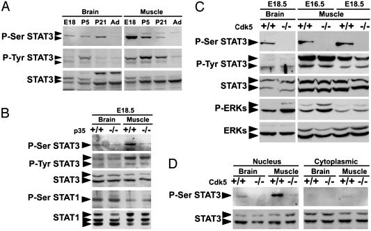
The activity of cyclin-dependent kinase 5 (Cdk5) depends on the association with one of its activators, p35 and p39, which are prominently expressed in the nervous system. Studies on the repertoire of protein substrates for Cdk5 have implicated the involvement of Cdk5 in neuronal migration and synaptic plasticity. Our recent analysis of the sequence of signal transducer and activator of transcription (STAT)3, a key transcription factor, reveals the presence of potential Cdk5 phosphorylation site. We report here that the Cdk5/p35 complex associates with STAT3 and phosphorylates STAT3 on the Ser-727 residue in vitro and in vivo. Intriguingly, whereas the Ser phosphorylation of STAT3 can be detected in embryonic and postnatal brain and muscle of wild-type mice, it is essentially absent from those of Cdk5-deficient embryos. In addition, treatment of cultured myotubes with neuregulin enhances the Ser phosphorylation of STAT3 and transcription of STAT3 target genes, such as c-fos and junB, in a Cdk5-dependent manner. Both the DNA-binding activity of STAT3 and the transcription of specific target genes, such as fibronectin, are reduced in Cdk5-deficient muscle. Taken together, these results reveal a physiological role of Cdk5 in regulating STAT3 phosphorylation and modulating its transcriptional activity.


Figures

Fig. 1.
Fig. 1.
Phosphorylation of STAT3 by active Cdk5 in vitro and in vivo. (A) Fusion protein of STAT3 C-terminal fragment (amino acids 660-770) was phosphorylated by recombinant Cdk5/p35 in vitro. Cdk5/p35 recombinant protein (0-1 μg per reaction as indicated) was added to the reaction mix. (B) Single mutation of STAT3 at Ser-727 (S727A) completely abolished the in vitro phosphorylation of STAT3 by Cdk5/p35 and Cdk5/p25. STAT3, wild-type STAT3 fragment; STAT3M, S727A mutant. (C) COS-7 cells were transfected with p35, dnCdk5/p35 or Cdk5/p35 expression constructs with (Right) or without STAT3 (Left). The ratio of DNAs used for transfection was indicated at the left at the top. V, mock transfected control. Total cell lysates were subjected to Western blot analysis by using P-Ser and P-Tyr STAT3-specific Abs. Total STAT3 and p35 served as controls. (D) p25 binds STAT3 in vitro. STAT3 overexpressed in COS-7 cells was pulled down by purified GST-p25 protein (1-5 μg), but not GST (5 μg). A weak interaction between STAT3 and GST-Cdk5 (5 μg) was also detected. (E) The C-terminal fragment of STAT3 (amino acids 660-770, 5 μg) interacts with p35 overexpressed in COS-7 cells. STAT3 and STAT3M showed similar affinity in their binding to p35. (F) Cdk5/p35 protein complex in E18 brain lysate was pulled down by GST-STAT3. (G) Association of p35 with STAT3 in COS-7 cells. The lysate from p35-overexpressing COS-7 cells was immunoprecipitated with p35 Ab and immunoblotted with Abs specific for STAT3 (Upper) or p35 (Lower). The rabbit normal IgG was used as a negative control. (H) Association of p35 with STAT3 in C2C12 myotubes and cortical neurons cultured for 21 days. Lysates of C2C12 myotubes or cortical neurons were immunoprecipitated by using p35 Ab and were immunoblotted with STAT3 Ab.
Fig. 2.
Fig. 2.
Attenuation of Ser phosphorylation of STAT3 in brain and muscle of mice lacking Cdk5 activity. (A) Developmental expression of phosphorylated and total STAT3 in rat brain and muscle. Western blot analysis of P-Ser STAT3, P-Tyr STAT3, and total STAT3 in rat brain and muscle are shown. P5, postnatal day 5; Ad, adult. (B) Western blot analysis of P-Ser and P-Tyr STAT3 and total STAT3 in brain and muscle of p35 wild-type (+/+) and mutant (-/-) embryos at E18.5. P-Ser STAT1, and total STAT1 served as control. (C) Western blot analysis of P-Ser STAT3, P-Tyr STAT3, and P-ERKs in brain and muscle of wild-type (+/+) and Cdk5 mutant (-/-) embryos at E16.5 and E18.5. Total STAT3 and ERK proteins served as control. (D) P-Ser and total STAT3 in the nuclear and cytoplasmic fractions in brain and muscle of wild-type (+/+) and Cdk5 mutant (-/-) embryos at E18.5.
Fig. 3.
Fig. 3.
Inhibition of Cdk5 activity abolished the NRG-induced Ser phosphorylation of STAT3. (A) Blockade of STAT3 activity and Cdk5 activity attenuated the NRG-induced c-fos transcription. Northern blot analysis of c-fos transcript after treatment of C2C12 myotubes with NRG (+) for 30 min. The myotubes were pretreated with different inhibitors: 40 μM Ros, 100 μM AG490 for 4 h, or 100 μM STAT3-inhibitory peptide (STAT3 peptide) for 24 h as indicated. (B) Cultured C2C12 myotubes were treated with NRG for 0-30 min. Total cell lysates were subjected to Western blot analysis using Abs specific for P-Ser STAT3, P-Tyr STAT3, and total STAT3 (Upper). Induction of P-ERKs served as positive control for NRG treatment (Lower). (C) Inhibition of Cdk5 activity with Ros abolished the NRG-induced P-Ser STAT3. -, untreated; +, NRG-treated for 15 min. DMSO served as control. (D) Expression of dnCdk5 in C2C12 myotubes attenuated the NRG-induced P-Ser STAT3. (E) Cultured C2C12 myotubes were treated with NRG for 0-30 min with or without Ros pretreatment. Lysates of nuclear (Left) or cytoplasmic (Right) fractions were prepared and were immunoblotted with specific Abs as depicted.
Fig. 4.
Fig. 4.
Cdk5 regulated the STAT3 DNA-binding and transcriptional activity in vitro and in vivo. (A) Blockade of Cdk5 activity by Ros inhibited the NRG-induced increase in DNA binding of STAT3 in C2C12 myotubes. (Lower) Total STAT3. (Right) DNA-binding activity of STAT3 α and β was quantitated and expressed as a ratio of that in control myotubes pretreated with DMSO. Each value represented the mean ± SEM. of three representative experiments; *, P < 0.005. (B) Northern blot analysis of c-fos and junB in primary muscle cultures prepared from E18.5 wild-type (+/+) or Cdk5 null (-/-) embryos with (+) or without (-) NRG treatment. (Upper) c-fos. (Lower) junB. (C) DNA binding of STAT3 in E18.5 Cdk5 wild-type (+/+) and mutant (-/-) muscle (Left) and quantitation (Right). DNA-binding activity of STAT3 α and β was quantitated and expressed as a ratio of that in wild-type muscle. Each value represented the mean ± SEM. of three representative experiments; *, P < 0.005. (D) Northern blot analysis of fibronectin and muscle creatine kinase (MCK) in wild-type (+/+) and Cdk5-deficient (-/-) muscle of E18.5 embryos. MCK served as an equal loading control. (E) Overexpression of p35 increased the transcriptional activity of STAT3 in primary chick myotubes in a dose-dependent manner. Primary chick myotubes were transiently transfected with a STAT3 (pSTAT3-TA-Luc) or STAT1 (pGAS-TA-Luc) reporter gene construct and an internal control plasmid (β-gal-pCMV) with or without p35 or dnCdk5. The ratio of DNAs used for transfection was as depicted on the x axis. Luciferase activity was measured and normalized against the β-gal activity in the samples. Promoter activity was expressed as the ratio of luciferase activity in cells transfected with different combinations of p35 and dnCdk5 relative to that transfected with the empty vector. The data represented the mean ± SEM, n = 5. (F) Overexpression of p35 increased the transcriptional activity of c-fos. c-fos promoter-luciferase assay was performed as described in E with pSTAT3-TA-Luc (pSTAT3) as a positive control. Preincubation with Ros reduced Cdk5/p35-mediated transcriptional activity of pSTAT3 or c-fos. Overexpression of p35 could not increase the luciferase reporter activity under the control of STAT1 enhancer-responsive element (pGAS). The data represented mean ± SEM, n = 3. (G) The increase of STAT3 transcriptional activity depends on Ser-727 phosphorylation of STAT3. Primary muscle cultures were transfected with indicated expression constructs and luciferase assay was performed as in E; mean ± SEM, n = 3.

References

    1. Dhavan, R. & Tsai, L.-H. (2001) Nat. Rev. Mol. Cell Biol. 2, 749-759. - PubMed
    1. Lew, J., Huang, Q. Q., Qi, Z., Winkfein, R. J., Aebersold, R., Hunt, T. & Wang, J. H. (1994) Nature 371, 423-426. - PubMed
    1. Tsai, L.-H., Delalle, I., Caviness, V. S., Jr., Chae, T. & Harlow, E. (1994) Nature 371, 419-423. - PubMed
    1. Tang, D., Yeung, J., Lee, K. Y., Matsushita, M., Matsui, H., Tomizawa, K., Hatase, O. & Wang, J. H. (1995) J. Biol. Chem. 270, 26897-26903. - PubMed
    1. Fu, A. K., Fu, W.-Y., Cheung, J., Tsim, K. W., Ip, F. C., Wang, J. H. & Ip, N. Y. (2001) Nat. Neurosci. 4, 374-381. - PubMed

Publication types