. 2004 Apr 27;101(17):6770-3.
doi: 10.1073/pnas.0401604101. Epub 2004 Apr 19.

A role of reactive oxygen species in apoptotic activation of volume-sensitive Cl(-) channel

Affiliations

A role of reactive oxygen species in apoptotic activation of volume-sensitive Cl(-) channel

Takahiro Shimizu et al. Proc Natl Acad Sci U S A. .

Abstract

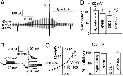
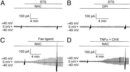
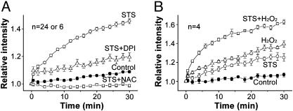
Apoptotic volume decrease is a pivotal event triggering a cell to undergo apoptosis and is induced by ionic effluxes resulting mainly from increased K(+) and Cl(-) conductances. Here, we demonstrate that in human epithelia HeLa cells both mitochondrion- and death receptor-mediated apoptosis inducers [staurosporine and Fas ligand or tumor necrosis factor (TNF)-alpha] rapidly activate Cl(-) currents that show properties phenotypical of volume-sensitive outwardly rectifying Cl(-) channel currents, including outward rectification, voltage-dependent inactivation gating at large positive potentials, inhibition by osmotic shrinkage, sensitivity to classic Cl(-) channel blockers, and dependence on cytosolic ATP. Staurosporine, but not Fas ligand or TNF-alpha, rapidly (within 30 min) increased the intracellular level of reactive oxygen species (ROS). A ROS scavenger and an NAD(P)H oxidase inhibitor blocked the current activation by staurosporine but not by Fas ligand or TNF-alpha. A ROS scavenger also inhibited apoptotic volume decrease, caspase-3 activation, and apoptotic cell death induced by staurosporine. Thus, it is concluded that an apoptosis-triggering anion conductance is carried by the volume-sensitive outwardly rectifying Cl(-) channel and that the channel activation on apoptotic stimulation with staurosporine, but not with Fas ligand or TNF-alpha, is mediated by ROS.


Figures

Fig. 1.
Fig. 1.
Activation of VSOR Cl- current by a mitochondrion-mediated apoptosis inducer, STS. (A) Representative record of whole-cell current before and during application of STS (4 μM), taken during application of alternating pulses from 0 to ±40 mV and, at selected time points, of step pulses from -100 to +100 mV in 20-mV increments. STS activated the current with a half activation time of 4.9 ± 0.5 min (n = 12). Note that the STS-induced current was inhibited by osmotic shrinkage induced by a hypertonic challenge. (B) Expanded traces of current responses to step pulses before (Left) and during (Right) stimulation with STS. Note that the STS-induced current exhibited faster inactivation kinetics at larger positive potentials. (C) I-V relationship for the mean densities of currents activated by STS. (D) Sensitivity of STS-induced current to shrinkage (induced by ≈5-min exposure to hypertonic solution of 116% osmolality), Cl- channel blockers (≈5-min treatment with 100 μM NPPB and 500 μM DIDS), and cytosolic ATP removal (>15-min equilibration with ATP-free pipette solution). The percent of inhibition was calculated from the peak current densities recorded at ±100 mV from the cells under control (isotonic, ATP-containing, and blocker-free) conditions (n = 27) and the cells under hypertonic, cytosolic ATP-free, or blocker-containing conditions. Sensitivity to DIDS was voltage-dependent, being significantly lower at -100 mV than at +100 mV.
Fig. 2.
Fig. 2.
Activation of VSOR Cl- current by receptor-mediated apoptosis inducers. Clamped voltages are the same as in Fig. 1 A. (A) Representative record of currents before and during application of anti-Fas antibody (500 ng/ml). Note the sensitivity of Fas ligand-induced current to osmotic shrinkage. (B) Representative record of currents before and during application of TNF-α (2 ng/ml) plus CHX (1 μg/ml). Note the sensitivity of TNF-α-induced current to NPPB (100 μM). (C and D) Sensitivity of currents activated by anti-Fas antibodies (C) and TNF-α+CHX (D) to osmotic shrinkage, NPPB, DIDS, and cytosolic ATP removal. The percent of inhibition was calculated by using the control data (n = 24 and 21 for C and D, respectively), as described in the legend for Fig. 1D. Significant voltage dependence was found only for sensitivity to DIDS.
Fig. 3.
Fig. 3.
Effects of a ROS scavenger (10 mM NAC in A, C, and D) and an NAD(P)H oxidase inhibitor (20 μM DPI in B) on STS-induced (A and B), Fas ligand-induced (C), or TNF-α-induced (D) activation of VSOR Cl- current. Clamped voltages are the same as in Fig. 1 A. Each trace represents five similar observations.
Fig. 4.
Fig. 4.
ROS production by STS. (A) Effects of NAC (10 mM) and DPI (20 μM) on STS-induced ROS production monitored by DCF fluorescence. Each symbol represents the mean ± SEM (bar) of 24 observations except for experiments with DPI (n = 6). STS significantly increased the level of intracellular ROS ≥0.5 min after stimulation. NAC and DPI significantly inhibited this increase observed 0.5-30 and 4-30 min, respectively, after STS stimulation. DPI or NAC alone did not affect the ROS level in the absence of STS (data not shown, n = 6 or 24). (B) Effects of H2O2 (500 μM) on the ROS level in the absence and presence of STS. STS, H2O2, and STS plus H2O2 significantly increased the level observed 6-30, 2-30, and 2-30 min, respectively, after application.
Fig. 5.
Fig. 5.
Activation of VSOR Cl- current by H2O2. (A) Representative record of whole-cell currents before and during application of H2O2 (500 μM). Clamped voltages are the same as in Fig. 1 A. Note the sensitivity of H2O2-induced current to osmotic shrinkage. The relative mean cell diameter compared to the control (without H2O2) was 0.99 ± 0.01 (n = 7; P > 0.05) 3 min before and 0.92 ± 0.02 (n = 4; P < 0.05) 5 min after a hypertonic challenge in the presence of H2O2. (B) Expanded traces of current responses to step pulses before (Left) and during (Right) stimulation with H2O2. Note the voltage-dependent inactivation kinetics of H2O2-induced current. (C) I-V relationship for the mean densities of currents activated by H2O2. (D) Sensitivity of the H2O2-induced current to osmotic shrinkage, NPPB, DIDS, and cytosolic ATP removal. The percent of inhibition was calculated by using the control data (n = 19), as described in the legend for Fig. 1D. Note the voltage-dependent sensitivity to DIDS.
Fig. 6.
Fig. 6.
Involvement of ROS in apoptotic events induced by STS. (A) Effects of NAC (10 mM) and DPI (20 μM) on induction of AVD by STS (4 μM). STS application caused significant decrease in the mean cell volume 1-30 min after stimulation with a half-maximum shrinkage time of 10.8 ± 0.7 min (n = 7). NAC and DPI significantly inhibited STS-induced AVD 2-30 and 5-30 min, respectively, after stimulation. NAC alone caused a significant increase in the mean cell volume, irrespective of STS stimulation. (B) Effects of H2O2 (500 μM) on mean cell volume and STS-induced AVD. H2O2 alone caused a significant decrease in the mean cell volume 10-30 min after stimulation. AVD induced by STS in the presence of H2O2 was of a significantly greater level than when H2O2 was absent, 5-15 min after stimulation. (C) Effects of NAC on activation of caspase-3 by 4-h application of STS. NAC significantly suppressed STS-induced caspase-3 activation. *, Significant difference between data with and without STS. †, Significant difference between data with and without NAC. (D) Effects of NAC on the induction of cell death by 8-h application of STS. Note the rescue from STS-induced cell death by NAC. *, Significantly different from the control cell viability.

References

    1. Okada, Y., Maeno, E., Shimizu, T., Dezaki, K., Wang, J. & Morishima, S. (2001) J. Physiol. (London) 532, 3-16. - PMC - PubMed
    1. Gomez-Angelats, M. & Cidlowski, J. A. (2002) Toxicol. Pathol. 30, 541-551. - PubMed
    1. Schumann, M. A., Gardner, P. & Raffin, T. A. (1993) J. Biol. Chem. 268, 2134-2140. - PubMed
    1. Szabo, I., Lepple-Wienhues, A., Kaba, K. N., Zoratti, M., Gulbins, E. & Lang, F. (1998) Proc. Natl. Acad. Sci. USA 95, 6169-6174. - PMC - PubMed
    1. Nietsch, H. H., Roe, M. W., Fiekers, J. F., Moore, A. L. & Lidofsky, S. D. (2000) J. Biol. Chem. 275, 20556-20561. - PubMed

Publication types