. 2004 Apr 27;101(17):6391-6.
doi: 10.1073/pnas.0401031101. Epub 2004 Apr 19.

Escherichia coli nucleoside diphosphate kinase does not act as a uracil-processing DNA repair nuclease

Affiliations

Escherichia coli nucleoside diphosphate kinase does not act as a uracil-processing DNA repair nuclease

Samuel E Bennett et al. Proc Natl Acad Sci U S A. .

Abstract

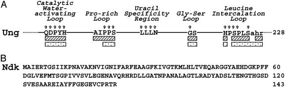
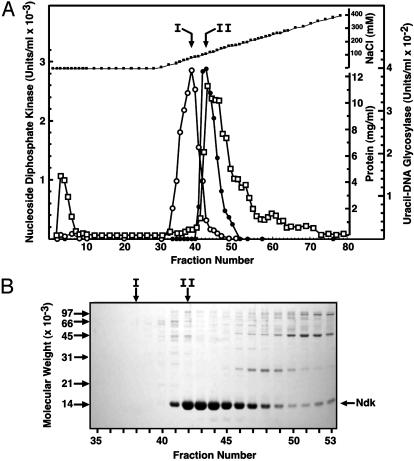
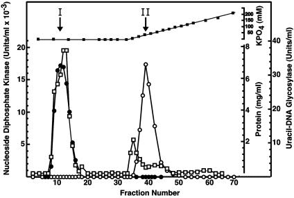
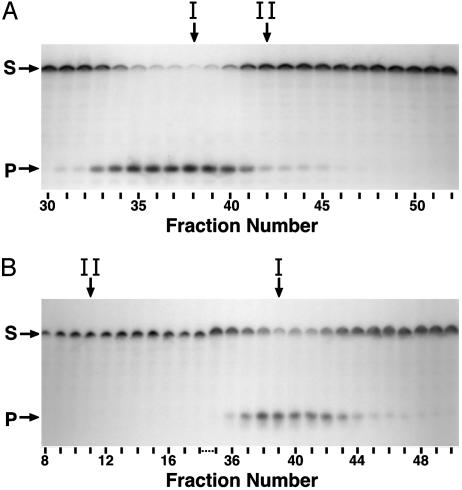
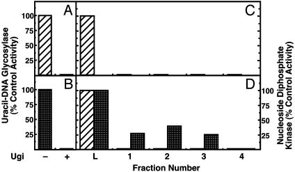
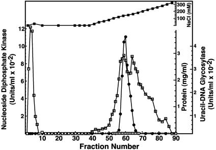
Escherichia coli nucleoside diphosphate kinase (Ndk) catalyzes ATP-dependent synthesis of ribo- and deoxyribonucleoside triphosphates from the cognate diphosphate precursor. Recently, the Ndk polypeptide was reported to be a multifunctional base excision repair nuclease that processed uracil residues in DNA by acting sequentially as a uracil-DNA glycosylase inhibitor protein (Ugi)-sensitive uracil-DNA glycosylase, an apurinic/apyrimidiniclyase, and a 3'-phosphodiesterase [Postel, E. H. & Abramczyk, B. M. (2003) Proc. Natl. Acad. Sci. USA 100, 13247-13252]. Here we demonstrate that the E. coli Ndk polypeptide lacked detectable uracil-DNA glycosylase activity and, hence, was incapable of acting as a uracil-processing DNA repair nuclease. This finding was based on the following observations: (i) uracil-DNA glycosylase activity did not copurify with Ndk activity; (ii) Ndk purified from E. coli ung(-) cells showed no detectable uracil-DNA glycosylase activity; and (iii) Ndk failed to bind to a Ugi-Sepharose affinity column that tightly bound E. coli uracil-DNA glycosylase (Ung). Collectively, these observations demonstrate that the E. coli Ndk polypeptide does not possess inherent uracil-DNA glycosylase activity.


Figures

Fig. 1.
Fig. 1.
Conserved motifs of E. coli Ung and amino acid sequence of E. coli Ndk. (A) Five structural motifs, previously implicated by Putnam et al. (18) as critical to Ung activity, are illustrated. Capitalized amino acids indicate structurally equivalent residues in E. coli, human, and herpes simplex virus uracil-DNA glycosylases as determined by high-resolution crystal structures (18, 34, 35). Starred residues correspond to amino acids that are absolutely conserved in E. coli, human, herpes simplex virus, and B. subtilis uracil-DNA glycosylase polypeptides. Striped and speckled bars correspond to residues that contact Ugi and DNA containing an AP-site, respectively. (B) The amino acid sequence of E. coli Ndk, as described by Hama et al. (2), is shown.
Fig. 2.
Fig. 2.
DEAE-Sepharose column elution profile of uracil-DNA glycosylase and Ndk activity. (A) DEAE-Sepharose column chromatography and standard assays for uracil-DNA glycosylase (open circles) and Ndk (filled circles) were performed as described in Materials and Methods. Protein concentration (open squares) and NaCl concentration (filled squares) were determined as described in Materials and Methods. (B) 12% SDS/PAGE of DEAE-Sepharose column fractions was conducted as described in Materials and Methods. The molecular weight standards are indicated by arrows on the left side of the gel; the location of the Ndk band is shown on the right side. Fractions corresponding to the peak of uracil-DNA glycosylase (I) and Ndk (II) activity are indicated by vertical arrows.
Fig. 3.
Fig. 3.
Hydroxyapatite column elution profile of E. coli Ung and Ndk activity. Hydroxyapatite column chromatography of Fraction III and standard assays for Ung (open circles) and Ndk (filled circles) were performed as described in Materials and Methods. Protein concentration (open squares) and potassium phosphate concentration (filled squares) were determined as described in Materials and Methods. Fractions corresponding to the peak of Ung (I) and Ndk (II) activity are indicated by vertical arrows.
Fig. 4.
Fig. 4.
Detection of uracil-DNA glycosylase activity by using a uracil-containing oligodeoxynucleotide substrate. Reaction mixtures (40 μl) containing 2 pmol of 5′-end 32P-labeled duplex oligonucleotide ([32P]U·A 21-mer) and 10 μl of various DEAE-Sepharose (A) or hydroxyapatite (B) fractions were prepared and processed as described in Materials and Methods. Samples (10 μl) of the reaction products were resolved by using denaturing 12% polyacrylamide/8.3 M urea gel electrophoresis. Arrows indicate the location on the autoradiogram of unreacted [32P]21-mer substrate (S) and [32P]9-mer products (P). Fractions corresponding to the peak of Ung (I) and Ndk (II) activity are indicated by vertical arrows.
Fig. 5.
Fig. 5.
Ugi-Sepharose column chromatography removes the uracil-DNA glycosylase activity from the Ndk preparation. Standard uracil-DNA glycosylase assays were performed with 1.3 units of E. coli Ung (hatched bars) (A) or 127 units of Ndk (Fraction IV, checkered bars) containing 0.16 units of Ung activity with (+) and without (-) 9 units of Ugi (B). The amount of uracil-DNA glycosylase activity detected in the absence of Ugi was set to 100%. (C) E. coli Ung (25 units in 125 μl, column L) was applied to a Ugi-Sepharose column (125 μl), and fractions were collected as described in Materials and Methods. Column fractions were assayed for uracil-DNA glycosylase activity (striped bars); the amount of uracil-DNA glycosylase activity present in the 125-μl sample (L) was set to 100%. (D) Ndk (Fraction IV containing 3.2 units of uracil-DNA glycosylase activity) was applied to a Ugi-Sepharose column and fractions were collected as described for C. Column fractions were assayed for uracil-DNA glycosylase activity (hatched bars) and Ndk activity (checkered bars). The amount of uracil-DNA glycosylase or Ndk activity present in the sample applied to column (L) was set to 100%.
Fig. 6.
Fig. 6.
DEAE-Sepharose column elution profile of E. coli Ndk activity over-produced in ung- E. coli Cells. DEAE-Sepharose column chromatography and standard assays for uracil-DNA glycosylase (open circles), Ndk (filled circles), protein concentration (open squares), and NaCl concentration (filled squares) were performed as described in Materials and Methods.

References

    1. Almaula, N., Lu, Q., Delgado, J. S. B. & Inouye, M. (1995) J. Bacteriol. 177, 2524-2529. - PMC - PubMed
    1. Hama, H., Almaula, N., Lerner, C. G., Inouye, S. & Inouye, M. (1991) Gene 105, 31-36. - PubMed
    1. Miller, J. H., Funchain, P., Clendenin, W., Huang, T., Nguyen, A., Wolff, E., Yeung, A., Chiang, J. H., Garibyan, L., Slupska, M. M. & Yang, H. (2002) Genetics 162, 5-13. - PMC - PubMed
    1. Lu, Q., Zhang, X., Almaula, N., Mathews, C. K. & Inouye, M. (1995) J. Mol. Biol. 254, 337-341. - PubMed
    1. Postel, E. H. & Abramczyk, B. M. (2003) Proc. Natl. Acad. Sci. USA 100, 13247-13252. - PMC - PubMed

Publication types

MeSH terms

LinkOut - more resources