. 2004 Apr 27;101(17):6792-7.
doi: 10.1073/pnas.0401090101. Epub 2004 Apr 19.

Arabidopsis thaliana has the enzymatic machinery for replicating representative viroid species of the family Pospiviroidae

Affiliations

Arabidopsis thaliana has the enzymatic machinery for replicating representative viroid species of the family Pospiviroidae

José-Antonio Daròs et al. Proc Natl Acad Sci U S A. .

Abstract

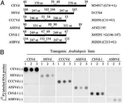
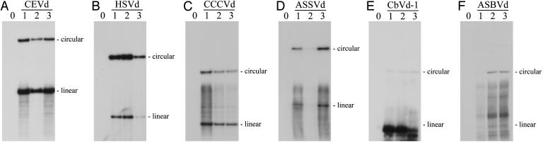
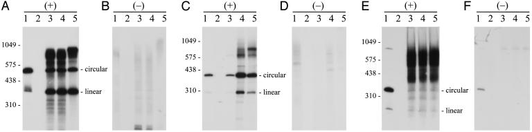
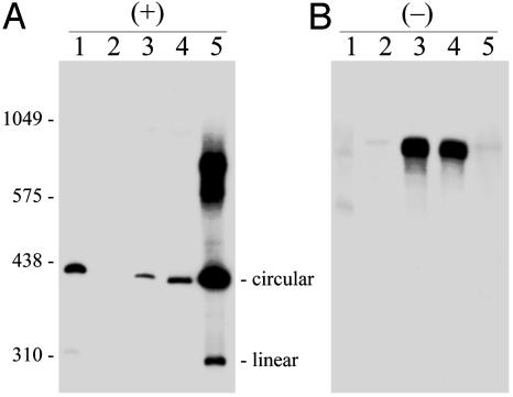
Viroids, subviral noncoding RNAs, replicate, move, and incite diseases in plants. Viroids replicate through a rolling-circle mechanism in which oligomeric RNAs of one or both polarities are cleaved and ligated into the circular monomers. Attempts to transmit viroids to Arabidopsis have failed for unknown reasons. To tackle this question, Arabidopsis was transformed with cDNAs expressing dimeric (+) transcripts of representative species of the families Pospiviroidae and Avsunviroidae, which replicate in the nucleus and the chloroplast, respectively. Correct processing to the circular (+) monomers was always observed, demonstrating that Arabidopsis has the appropriate RNase and RNA ligase. Northern blot hybridization also revealed the multimeric (-) RNAs of Citrus exocortis viroid and Hop stunt viroid (HSVd) of the family Pospiviroidae, but not of Avocado sunblotch viroid of the family Avsunviroidae, showing that the first RNA-RNA transcription of the rolling-circle mechanism occurs in Arabidopsis for the two nuclear viroids and that their multimeric (-) RNAs remain unprocessed as in typical hosts. Moreover, transgenic Arabidopsis expressing HSVd dimeric (-) transcripts accumulated the circular (+) monomers, although at low levels, together with the unprocessed primary transcript that served as the template for the second RNA-RNA transcription. Agroinoculation of Arabidopsis with the dimeric (+) Citrus exocortis viroid, HSVd, and Coleus blumei viroid 1 cDNAs showed that these viroids could not move to distal plant parts, in contrast with the situation observed in their experimental hosts. Therefore, deficiencies in movement or low replication appear to be the factors limiting infectivity of some viroids in Arabidopsis.


Figures

Fig. 1.
Fig. 1.
(A) Bar diagram representing the viroid sequences embedded in the transcripts expressed in the transgenic Arabidopsis. Viroid species are indicated on the left, and the database accession numbers of the particular sequence variants used are on the right. Whenever sequence variants do not exactly match a database entry, the differences are indicated in parentheses after the accession number. Positions where the viroid sequences start and end in the transcript and the size of each monomeric unit are also indicated. (B) Dot-blot hybridization analysis of transgenic Arabidopsis expressing different dimeric (+) viroid RNAs. Total RNAs from three independent transgenic lines (columns 1, 2, and 3) for each of the six constructs (CEVd, HSVd, CCCVd, ASSVd, CbVd-1, and ASBVd) were hybridized with each of the six 32P-labeled complementary riboprobes indicated on the left.
Fig. 2.
Fig. 2.
Northern blot hybridization analysis of transgenic Arabidopsis expressing different dimeric (+) viroid RNAs. Nucleic acid preparations from a nontranformed Arabidopsis control (lane 0) and three independent transgenic lines (lanes 1, 2, and 3) expressing dimeric (+) RNAs of CEVd (A), HSVd (B), CCCVd (C), ASSVd (D), CbVd-1 (E), and ASBVd (F) were separated by two consecutive PAGE steps. A segment of the first nondenaturing gel, which included the RNAs with sizes between ≈200 and ≈400 nucleotides, was cut and applied on top of the second denaturing gel. After separation in this second gel, RNAs were blotted to nylon membranes and hybridized with 32P-labeled riboprobes complementary to each of the viroid RNAs. The analysis was performed with total RNA preparations except for CbVd-1, in which a viroid-enriched preparation obtained by chromatography on nonionic cellulose was used. Bands with the mobilities of the corresponding monomeric circular and linear viroid RNAs are indicated on the right (positions of linear CbVd-1 and ASBVd forms are only tentative). The exposure time was not the same in all autoradiographs.
Fig. 3.
Fig. 3.
Northern blot hybridization analysis of transgenic Arabidopsis expressing different dimeric (+) viroid RNAs. Viroid-enriched RNA preparations, obtained by chromatography on nonionic cellulose, were fractionated by single denaturing PAGE in duplicated gels and blotted to nylon membranes that were hybridized with 32P-labeled riboprobes for detecting the (+) and (-) strands of CEVd (A and B), HSVd (C and D), and ASBVd (E and F), respectively. Lanes 1, controls of CEVd-infected gynura (A and B), HSVd-infected cucumber (C and D), and ASBVd-infected avocado (E and F); lanes 2, nontransformed Arabidopsis control; lanes 3-5, transgenic Arabidopsis expressing dimeric (+) RNAs of CEVd (A and B), HSVd (C and D), and ASBVd (E and F). Positions of linear RNA markers, with their size in nucleotides, are indicated on the left in A, C, and E. Positions of circular and linear CEVd, HSVd, and ASBVd monomeric RNAs are indicated on the right in A, C, and E, respectively. Both riboprobes for each viroid were equalized in acid-precipitable counts, and the films were exposed for the same time. For facilitating detection of CEVd and HSVd (-) strands, the volume of applied extract was 10-fold higher.
Fig. 4.
Fig. 4.
Northern blot hybridization analysis of transgenic Arabidopsis expressing dimeric (+) and (-) HSVd RNAs. Viroid-enriched RNA preparations, obtained by chromatography on nonionic cellulose, were fractionated by single denaturing PAGE in duplicated gels and blotted to nylon membranes that were hybridized with 32P-labeled riboprobes for detecting the (+) and (-) HSVd strands (A and B, respectively). Lanes 1, control of HSVd-infected cucumber; lanes 2, control of nontransformed Arabidopsis; lanes 3 and 4, two independent transgenic Arabidopsis expressing dimeric (-) HSVd RNA; and lanes 5, transgenic Arabidopsis expressing dimeric (+) HSVd RNA. Positions of linear RNA markers, with their size in nucleotides, are indicated on the left. Positions of circular and linear HSVd RNAs are indicated on the right in A. Both riboprobes were equalized in acid-precipitable counts, and the films were exposed for the same time. For facilitating detection of HSVd strands accumulating at lower levels, the volume of applied extract was 10-fold higher in lanes 3 and 4 in A, and in lanes 1 and 5 in B.

References

    1. Somerville, C. & Koornneef, M. (2002) Nat. Rev. Genet. 3, 883-889. - PubMed
    1. Arabidopsis Genomic Initiative. (2000) Nature 408, 796-815. - PubMed
    1. Yamanaka, T., Ohta, T., Takahashi, M., Meshi, T., Schmidt, R., Dean, C., Naito, S. & Ishikawa, M. (2000) Proc. Natl. Acad. Sci. USA 97, 10107-10112. - PMC - PubMed
    1. Lellis, A. D., Kasschau, K. D., Whitham, S. A. & Carrington, J. C. (2002) Curr. Biol. 25, 1046-1051. - PubMed
    1. Chisholm, S. T., Mahajan, S. K., Whitham, S. A., Yamamoto, M. L. & Carrington, J. C. (2000) Proc. Natl. Acad. Sci. USA 97, 489-494. - PMC - PubMed

Publication types

LinkOut - more resources