. 2004 Apr 27;101(17):6698-703.
doi: 10.1073/pnas.0303859101. Epub 2004 Apr 19.

Fibrin depletion decreases inflammation and delays the onset of demyelination in a tumor necrosis factor transgenic mouse model for multiple sclerosis

Affiliations

Fibrin depletion decreases inflammation and delays the onset of demyelination in a tumor necrosis factor transgenic mouse model for multiple sclerosis

Katerina Akassoglou et al. Proc Natl Acad Sci U S A. .

Abstract

In multiple sclerosis, in which brain tissue becomes permeable to blood proteins, extravascular fibrin deposition correlates with sites of inflammatory demyelination and axonal damage. To examine the role of fibrin in neuroinflammatory demyelination, we depleted fibrin in two tumor necrosis factor transgenic mouse models of multiple sclerosis, transgenic lines TgK21 and Tg6074. In a genetic analysis, we crossed TgK21 mice into a fibrin-deficient background. TgK21fib(-/-) mice had decreased inflammation and expression of major histocompatibility complex class I antigens, reduced demyelination, and a lengthened lifespan compared with TgK21 mice. In a pharmacologic analysis, fibrin depletion, by using the snake venom ancrod, in Tg6074 mice also delayed the onset of inflammatory demyelination. Overall, these results indicate that fibrin regulates the inflammatory response in neuroinflammatory diseases. Design of therapeutic strategies based on fibrin depletion could potentially benefit the clinical course of demyelinating diseases such as multiple sclerosis.


Figures

Fig. 1.
Fig. 1.
Inflammatory demyelinating lesions of TgK21 mice have fibrin deposition but normal amounts of plasminogen activator activity. Luxol fast blue staining counterstained with nuclear red shows normal histology in WT mice (A), whereas TgK21 spinal cords have meningeal inflammation (arrows) and myelin loss (asterisk) (B). Immunostaining for fibrin(ogen) shows that WT mice do not have fibrinogen in their spinal cord (C), whereas TgK21 spinal cords are immunoreactive for fibrin(ogen) (D). In situ zymography on spinal cord sections shows proteolytic activity in both normal (E) and TgK21 (F) mice. (Bar = 722 μm for A-D, 1.6 mm for E and F.)
Fig. 2.
Fig. 2.
Fibrin deposition precedes demyelination and correlates with demyelinating plaques in TgK21 mice. Immunohistochemistry for fibrin(ogen) shows deposition of fibrin (A) at areas of meningeal inflammation before the onset of demyelination (B). In addition, fibrin immunoreactivity (C) is detected at areas the spinal cord associated with inflammation and loss of myelin (D). A and C and B and D represent high-magnification images of Fig. 1 D and B, respectively. (Bar = 125 μm.)
Fig. 3.
Fig. 3.
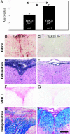
Fibrin(ogen) facilitates inflammatory demyelination. (A) TgK21fib-/- demonstrate a 1.6-fold lifespan increase (6.4 ± 0.4 weeks; n = 11) compared with TgK21fib+/+ littermate mice (4.2 ± 0.3 weeks; n = 9, P < 0.01). Immunohistochemistry for fibrin shows fibrin deposition in TgK21 spinal cord (B), but fibrin is absent from TgK21fib-/- spinal cord (C). Hematoxylin/eosin staining shows accumulation of inflammatory cells in TgK21 spinal cord (D), whereas there is minor inflammation in TgK21fib-/- spinal cord (E). Immunohistochemistry for MHC class I shows the presence of cytotoxic cells in TgK21 (F), which are absent from TgK21fib-/- (G). Myelin staining with luxol fast blue (blue) counterstained with periodic acid-Schiff stain (phagocytic macrophages, purple) shows active demyelination in TgK21 (H), whereas there are no lesions in TgK21fib-/- (I). (Bar = 170 μm.)
Fig. 4.
Fig. 4.
Pharmacologic depletion of fibrin(ogen) delays the onset of inflammatory demyelination. Tg6074 transgenic mice show myelin loss at the cerebellum by 6 weeks of age (A, arrows), whereas Tg6074 mice pretreated with ancrod for 2 weeks show no myelin alteration by 6 weeks of age (B). Tg6074 mice show up-regulation of MHC class I genes in the cerebellum (C), whereas fibrin-depleted Tg6074 do not show any signs of inflammatory cells or expression of immunologic markers characteristic of cytotoxic immune response (D). (Bar = 200 μm.)
Fig. 5.
Fig. 5.
Fibrinogen induces differentiation in macrophages. Untreated CD11b-immunostained RAW 264.7 macrophage cells (A) show undifferentiated, macrophage-like morphology. LPS- (1 μg/ml; B) or fibrinogen- (50 μg/ml; C) stimulated RAW264.7 cells show a morphological transformation to dendritic-like cells. LPS-stimulated cells (E) show morphologic alterations similar to those of fibrinogen-stimulated cells (F). Untreated cells (D) are round and smaller. (G) Quantitation of cells with activated morphology shows a statistically significant increase in activation upon fibrinogen stimulation, compared with untreated or fibronectin-stimulated cells. (H) Proliferation assay shows a statistically significant decrease of macrophage proliferation upon fibrinogen induction, compared with untreated or fibronectin-stimulated cells. Results are presented as means ± SE. Statistical analysis was performed by using Student's t test. (Scale bar = 45μm for A-C, 25 μm for D-F.)

Similar articles

Cited by

References

    1. Furie, B. & Furie, B. C. (1988) Cell 53, 505-518. - PubMed
    1. Colvin, R. B., Johnson, R. A., Mihm, M. C., Jr., & Dvorak, H. F. (1973) J. Exp. Med. 138, 686-698. - PMC - PubMed
    1. Levy, G. A., Leibowitz, J. L. & Edgington, T. S. (1981) J. Exp. Med. 154, 1150-1163. - PMC - PubMed
    1. Neale, T. J., Tipping, P. G., Carson, S. D. & Holdsworth, S. R. (1988) Lancet 2, 421-424. - PubMed
    1. Labarrere, C. A., Nelson, D. R. & Faulk, W. P. (1998) Am. J. Med. 105, 207-213. - PubMed

Publication types