. 2004 May;13(5):1331-9.
doi: 10.1110/ps.04618904.

An efficient system for high-level expression and easy purification of authentic recombinant proteins

Affiliations

An efficient system for high-level expression and easy purification of authentic recombinant proteins

Ann-Maree Catanzariti et al. Protein Sci. 2004 May.

Abstract

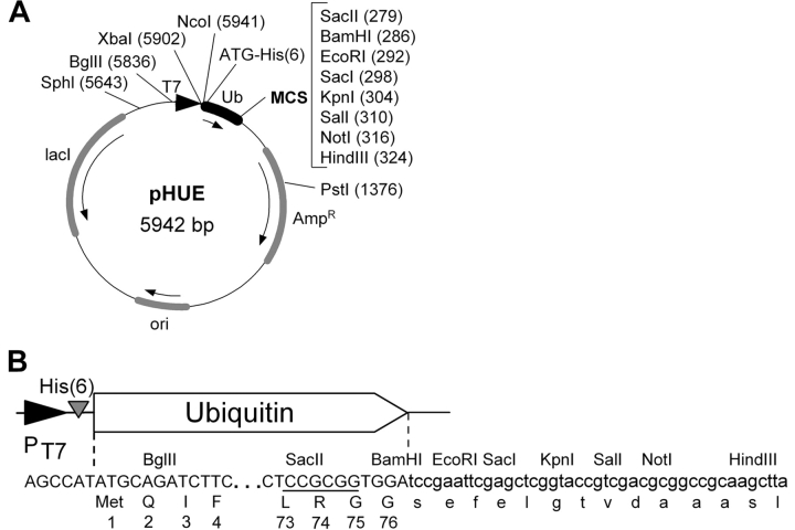
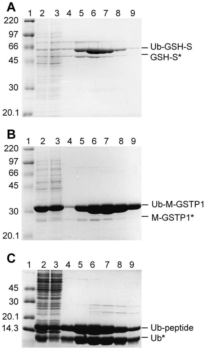
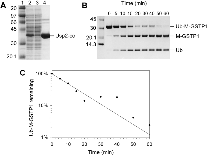
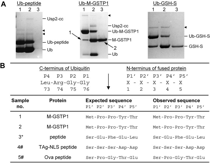
Expression of recombinant proteins as fusions to the eukaryotic protein ubiquitin has been found to significantly increase the yield of unstable or poorly expressed proteins. The benefit of this technique is further enhanced by the availability of naturally occurring deubiquitylating enzymes, which remove ubiquitin from the fusion product. However, the versatility of the system has been constrained due to the lack of a robust, easily purified deubiquitylating enzyme. Here we report the development of an efficient expression system, utilizing the ubiquitin fusion technique, which allows convenient high yield and easy purification of authentic protein. An Escherichia coli vector (pHUE) was constructed for the expression of proteins as histidine-tagged ubiquitin fusions, and a histidine-tagged deubiquitylating enzyme to cleave these fusions was expressed and purified. The expression system was tested using several proteins varying in size and complexity. These results indicate that this procedure will be suitable for the expression and rapid purification of a broad range of proteins and peptides, and should be amenable to high-throughput applications.


Figures

Figure 1.
Figure 1.
The Histidine-tagged Ubiquitin Expression vector, pHUE. (A) Plasmid map of pHUE showing the ubiquitin (Ub) coding region (black box), the T7 polymerase promoter (black triangle), and other regions (shaded boxes). Arrows indicate the direction of transcription. Restriction enzyme recognition sites within the multiple cloning site (MCS) are listed and other useful recognition sites in the vector backbone are also shown (unique, except BglII); locations are given relative to the start codon upstream of the his-tag, ATG = 1. (His)6, poly histidine tag; Ampr, β-lactamase gene; ori, colE1 origin of replication; lacI, lacI repressor gene. (B) DNA and encoded protein sequence of the 5′ and 3′ end of the ubiquitin coding region showing the engineered SacII site (underlined) within codons Leu 73, Arg 74, and Gly 75, and the 3′ polylinker. Restriction sites and protein translation are given above and under the DNA sequence, respectively.
Figure 2.
Figure 2.
Purification of His-tagged Ub fusion proteins. Ub fusion proteins were isolated from crude E. coli extract by Ni-affinity chromatography under native conditions. Samples from sequential steps in the purification were resolved by 10% SDS-PAGE and stained with Coomassie blue. (A) Ub–GSH-S; (B) Ub–M-GST; (C) Ub-peptide. Molecular mass marker (lane 1); crude E. coli extract (lane 2); unbound proteins (lane 3); and elutions with 50 mM (lane 4), 100 mM (lane 5), 150 mM (lane 6), 200 mM (lane 7), or 250 mM (lanes 8,9) imidazole. Proteins migrating at the expected molecular mass of the Ub fusion are indicated on the right. Proteins with an asterisk represent apparent cleavage by an unknown E. coli protease (see text).
Figure 3.
Figure 3.
Purification of His-tagged Usp2-cc and time course cleavage assay. (A) Purification of His6Usp2-cc by Ni-affinity chromatography under native conditions. Samples from sequential steps in the purification were resolved by 10% SDS-PAGE and stained with Coomassie blue. (Lane 1) Marker (masses shown at left); (lane 2) crude E. coli extract; (lane 3) unbound proteins; (lane 4) elution with 150 mM imidazole. The protein migrating with the expected molecular mass of poly-his-tagged Usp2-cc is indicated on the right. (B) Purified Usp2-cc was assayed against the Ub-M-GSTP1 test substrate over a 60-min time course at a 1 : 100 molar ratio. Samples were taken at 0, 5, 10, 15, 20, 30, 40, 50, and 60 min (shown above lanes), then resolved by 10% SDS-PAGE and Coomassie blue staining. The position of Ub–M-GSTP1 and the two cleaved products, M-GSTP1 and Ub, are indicated on the right. (C) Densitometry was used to quantify the bands from panel B, and plotted as percent uncleaved substrate remaining (on a natural log scale) over time. Values were normalized for background pixelation and loading errors. The initial rate was found to be 0.4 μg Ub–M-GSTP1 cleaved per minute at this 1 : 100 enzyme : substrate ratio. This equates to a rate of 6.3 mg Ub–M-GSTP1 cleaved per minute per milligram enzyme.
Figure 4.
Figure 4.
Purification and N-terminal sequencing of cleaved proteins. (A) The Ub fusion proteins named above the gels were cleaved with Usp2-cc, then purified from the reaction mix by nickel-affinity chromatography. For each substrate, samples were taken before cleavage (lane 1), after cleavage (lane 2), and the supernatant after purification (lane 3). Proteins were resolved by SDS-PAGE and Coomassie blue staining in a 10% Tricine gel. Protein positions are indicated on the right. Black arrowheads indicate contaminating proteins. Numbered arrows show protein bands excised from the gel for N-terminal sequencing in panel B. (B) N-terminal sequencing of Usp2-cc cleaved and apparent E. coli-cleaved products. (Upper) The expected site of Usp2-cc cleavage is shown (↓). (Lower) Sample numbers correspond to the protein band excised from the SDS-polyacrylamide gel shown in panel A or data not shown. (*) Supernatant after purification of Ub-peptide cleavage reaction (panel A, lane 3, Ub-peptide gel). (#) Following HPLC purification of cleavage reaction supernatant.
Figure 5.
Figure 5.
Peptides produced as ubiquitin fusions retain activity. An ELISA-based binding assay was performed using microtiter plates coated with the protein or peptide indicated, and the change in absorbance (405 nm) plotted against concentration of Importin α/β heterodimer used. Apparent dissociation constants (Kd) were determined following curve-fitting (see Materials and Methods). (A) Data not corrected for His6-Ub binding; (B) data corrected for His6-Ub binding; (C) data obtained from a synthetic T-ag NLS peptide.

Similar articles

Cited by

References

    1. Bachmair, A., Finley, D., and Varshavsky, A. 1986. In vivo half-life of a protein is a function of its amino-terminal residue. Science 234 179–186. - PubMed
    1. Baker, R.T. 1996. Protein expression using ubiquitin fusion and cleavage. Curr. Opin. Biotechnol. 7 541–546. - PubMed
    1. Baker, R.T. and Board, P.G. 1991. The human ubiquitin-52 amino acid fusion protein gene shares several structural features with mammalian ribosomal protein genes. Nucleic Acids Res. 19 1035–1040. - PMC - PubMed
    1. Baker, R.T. and Varshavsky, A. 1995. Yeast N-terminal amidase. A new enzyme and component of the N-end rule pathway. J. Biol. Chem. 270 12065–12074. - PubMed
    1. Baker, R.T., Smith, S.A., Marano, R., McKee, J., and Board, P.G. 1994. Protein expression using cotranslational fusion and cleavage of ubiquitin. Mutagenesis of the glutathione-binding site of human Pi class glutathione S-transferase. J. Biol. Chem. 269 25381–25386. - PubMed

Publication types

MeSH terms

Associated data

LinkOut - more resources