. 2004;125(3):759-67.
doi: 10.1016/j.neuroscience.2004.01.049.

Chronic stress enhances ibotenic acid-induced damage selectively within the hippocampal CA3 region of male, but not female rats

Affiliations

Chronic stress enhances ibotenic acid-induced damage selectively within the hippocampal CA3 region of male, but not female rats

C D Conrad et al. Neuroscience. 2004.

Abstract

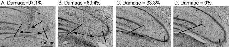
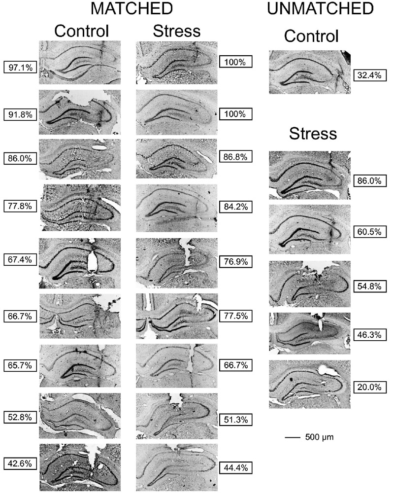
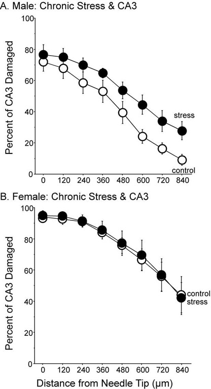
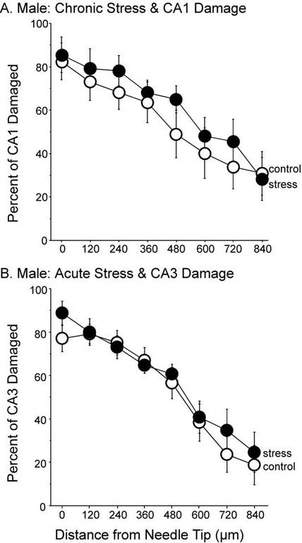
The purpose of this investigation was to assess the ability of the hippocampus to withstand a metabolic challenge following chronic stress. An N-methyl-d-aspartate receptor excitotoxin (ibotenic acid, IBO) was infused into the CA3 region of the hippocampus following a period of restraint for 6 h/day/21 days. Following the end of restraint when CA3 dendritic retraction persists (3 to 4 days), rats were infused with IBO (or vehicle) into the CA3 region of the hippocampus. Stressed male rats showed significantly more CA3 damage after IBO infusion relative to controls and the saline-infused side. Moreover, IBO-exacerbation of damage in males was not observed in the CA3 region 3 to 4 days after acute stress (6 h restraint), nor in the CA1 region after chronic stress. Females were also examined and chronic stress did not exacerbate IBO damage in the CA3 region. Overall, these results demonstrate that chronic stress compromises the ability of the hippocampus to withstand a metabolic challenge days after the chronic stress regimen has subsided in male rats. Whether the conditions surrounding CA3 dendritic retraction in females represents vulnerability is less clear and warrants further investigation.


Figures

Fig. 1
Fig. 1
Example of quantification of IBO-induced CA3 damage. The section containing the needle tip was identified (A). The single-headed arrow points to the region of the needle tip, indicated by pronounced gliosis. This example represents an ideal needle placement and corresponds to the first control rat in Fig. 2. The percent CA3 damage was determined by quantifying the length of CA3 that lacked pyramidal cells, indicated by the double-headed arrow. This value was divided by the total length of the CA3 region that previously expressed stress-induced dendritic retraction, which is indicated by the two slashed lines. In panel A, this region represents 97.1% damage. As the distance from the needle tip increases, the percent of CA3 damage decreases (B=69.4%, C=33.3%) until no damage is detected, D=0%. Magnification=80×.
Fig. 2
Fig. 2
A histological summary of IBO-induced CA3 damage in male rats. After determining the section containing the needle tip, the amount of damage in the CA3 region was determined without knowledge of treatment group. Rats were matched based upon similar needle-tip locations and CA3 damage. CA3 damage had to be greater than 20% and be within a 10% range of the other treatment to be matched. Nine pairings were made in ascending order of percent CA3 damage with the greatest CA3 damage illustrated by the first pair and the least CA3 damage illustrated by the ninth pair. Brains that were not matched are represented in the far right column. The boxes indicate the percent of damage in the section containing the needle tip.
Fig. 3
Fig. 3
Percent of CA3 damage after IBO infusion and chronic stress. (A) Males with a history of chronic stress expressed more IBO-induced CA3 damage compared to controls, F(1,16)=5.308, P<0.05. Damage in the section containing the needle tip did not differ among groups. Nine subjects per group. (B) In females, a history of chronic stress did not alter IBO-induced damage in the CA3 region, F(1,14)=0.017, P=0.899. Eight subjects per group. Data represent means±S.E.M.
Fig. 4
Fig. 4
(A) Percent of CA1 damage after IBO infusion in male rats exposed to chronic stress. A history of chronic stress had no influence on the amount of damage inflicted in the CA1 region after IBO infusion, F(1,12)=0.434, P=0.522. Seven subjects per group. (B) Percent of CA3 damage after IBO infusion in male rats exposed to acute stress. A single session of restraint did not alter IBO-induced CA3 damage, F(1,10)=0.139, P=0.717. Six subjects per group. Data represent means±S.E.M.

References

    1. Baran SE, Wright RL, Jackson JL, Kleen JK, Tsekhanov S, Wise L, Zachow KA, Conrad CD. Ovariectomized female rats demonstrate enhanced spatial memory on the Y-maze following chronic stress while acute estrogen treatment may attenuate performance. Soc Neurosci Abst. 2002;28:370.12.
    1. Cohen J, Cohen P, West SG, Aiken LS. Applied multiple regression/correlation analysis for the behavioral sciences. 3rd Lawrence Erlbaum Associates, Inc; Mahwah, New Jersey: 2003.
    1. Conrad CD, Galea LAM, Kuroda Y, McEwen BS. Chronic stress impairs rat spatial memory on the Y-Maze and this effect is blocked by tianeptine pre-treatment. Behav Neurosci. 1996;110:1321–1334. - PubMed
    1. Conrad CD, Magariños AM, LeDoux JE, McEwen BS. Repeated restraint stress increases fear conditioning, independently of causing hippocampal CA3 dendritic atrophy. Behav Neurosci. 1999;113:902–913. - PubMed
    1. Conrad CD, Mauldin-Jourdain ML, Hobbs RJ. Metryapone reveals that previous chronic stress differentially impairs hippocampal-dependent memory. Stress. 2001;4:305–318. - PMC - PubMed

Publication types

MeSH terms

LinkOut - more resources