

Domain insertions in protein structures
- PMID: 15099733
- PMCID: PMC2665287
- DOI: 10.1016/j.jmb.2004.03.039
Domain insertions in protein structures
Abstract
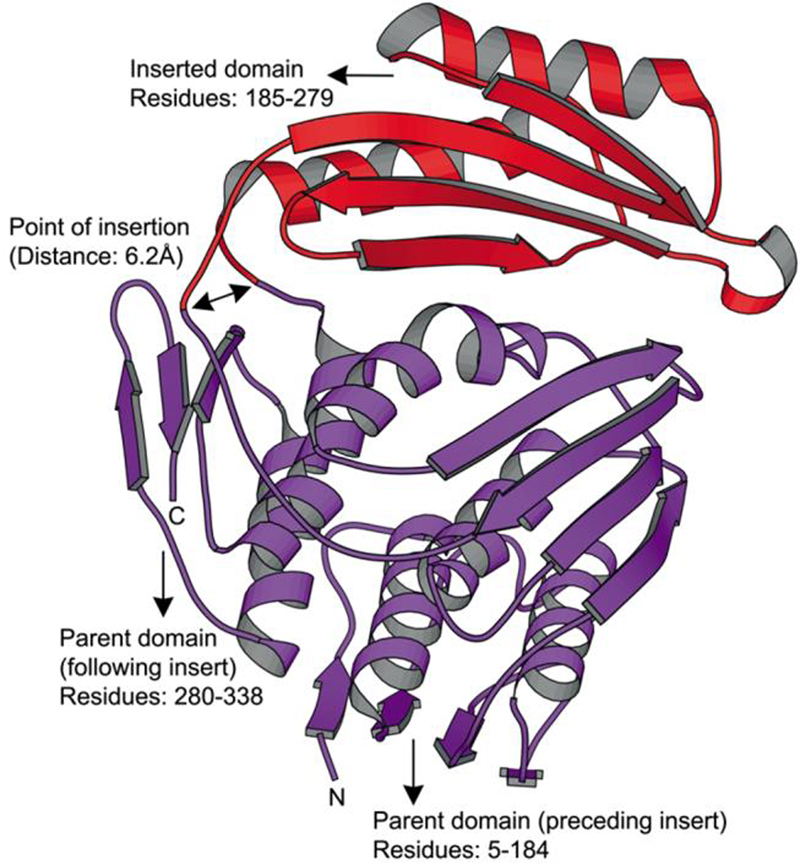
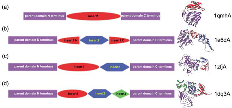
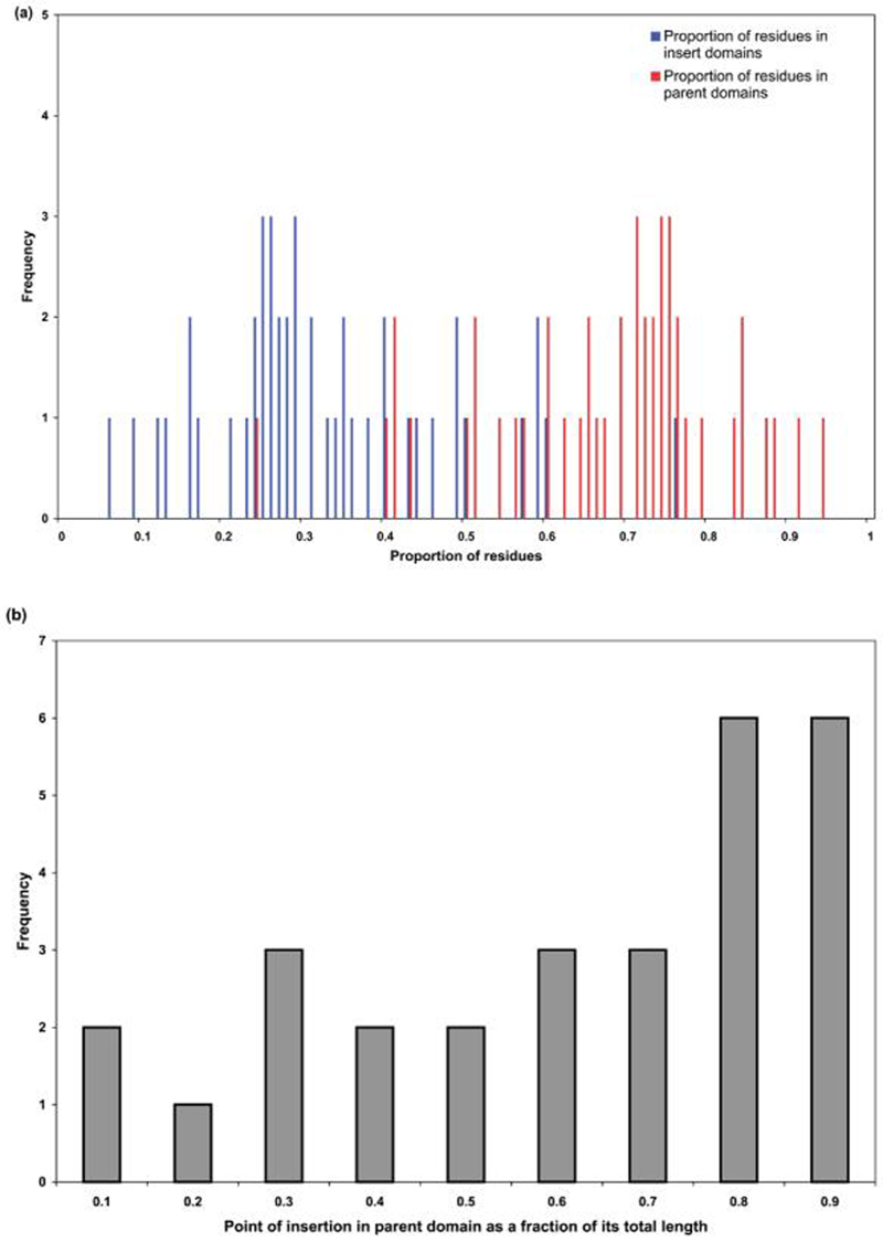
Domains are the structural, functional or evolutionary units of proteins. Proteins can comprise a single domain or a combination of domains. In multi-domain proteins, the domains almost always occur end-to-end, i.e., one domain follows the C-terminal end of another domain. However, there are exceptions to this common pattern, where multi-domain proteins are formed by insertion of one domain (insert) into another domain (parent). Here, we provide a quantitative description of known insertions in the Protein Data Bank (PDB). We found that 9% of domain combinations observed in non-redundant PDB are insertions. Although 90% of all insertions involve only one insert, proteins can clearly have multiple (nested, two-domain and three-domain) inserts. We also observed correlations between the structure and function of a domain and its tendency to be found as a parent or an insert. There is a bias in insert position towards the C terminus of parents. We observed that the atomic distance between the N and C terminus of an insert is significantly smaller when compared to the N-to-C distance in a parent context or a single domain context. Insertions are found always to occur in loop regions of parent domains. Our observations regarding the relationship between domain insertions and the structure, function and evolution of proteins have implications for protein engineering.
Figures
References
-
- Murzin AG, Brenner SE, Hubbard T, Chothia C. SCOP: a structural classification of proteins database for the investigation of sequences and structures. J. Mol. Biol. 1995;247:536–540. - PubMed
-
- Orengo CA, Michie AD, Jones S, Jones DT, Swindells MB, Thornton JM. CATH—a hierarchic classification of protein domain structures. Structure. 1997;5:1093–1108. - PubMed
-
- Holm L, Sander C. Mapping the protein universe. Science. 1996;273:595–603. - PubMed
-
- Chothia C. Proteins. One thousand families for the molecular biologist. Nature. 1992;357:543–544. - PubMed
-
- Bork P, Downing AK, Kieffer B, Campbell ID. Structure and distribution of modules in extracellular proteins. Quart. Rev. Biophys. 1996;29:119–167. - PubMed
Publication types
MeSH terms
LinkOut - more resources
Full Text Sources
Other Literature Sources
