. 2004 May 1;172(9):5287-96.
doi: 10.4049/jimmunol.172.9.5287.

Distinct IL-2 receptor signaling pattern in CD4+CD25+ regulatory T cells

Affiliations

Distinct IL-2 receptor signaling pattern in CD4+CD25+ regulatory T cells

Steven J Bensinger et al. J Immunol. .

Abstract

Despite expression of the high-affinity IL-2R, CD4(+)CD25(+) regulatory T cells (Tregs) are hypoproliferative upon IL-2R stimulation in vitro. However the mechanisms by which CD4(+)CD25(+) T cells respond to IL-2 signals are undefined. In this report, we examine the cellular and molecular responses of CD4(+)CD25(+) Tregs to IL-2. IL-2R stimulation results in a G(1) cell cycle arrest, cellular enlargement and increased cellular survival of CD4(+)CD25(+) T cells. We find a distinct pattern of IL-2R signaling in which the Janus kinase/STAT pathway remains intact, whereas IL-2 does not activate downstream targets of phosphatidylinositol 3-kinase. Negative regulation of phosphatidylinositol 3-kinase signaling and IL-2-mediated proliferation of CD4(+)CD25(+) T cells is inversely associated with expression of the phosphatase and tensin homologue deleted on chromosome 10, PTEN.


Figures

FIGURE 1
FIGURE 1
IL-2 mediates the survival and cellular enlargment of CD4+CD25+ T cells in vitro. CD4+CD25+ and CD4+CD25 cells were sorted by flow cytometry, CFSE labeled and cultured in complete medium alone or with increasing concentrations of IL-2 as indicated for 96 h. A, B, and D, Cells were stained with the vital dye 7-AAD or fixed permeabilized and stained with anti-Bcl-2 before analysis by flow cytometry. Results are representative of five independent experiments. C, Cells cultured as in A were lysed and electrophoresed on a SDS-PAGE gel, transferred to nitrocellulose membranes, and probed with anti-bcl-xL, or anti-actin as indicated. Primed CD4+ T cells stimulated with anti-CD3 for 96 h were used as a positive control for bcl-xL expression. Results are representative of four independent experiments.
FIGURE 2
FIGURE 2
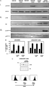
CD4+CD25+ T cells have distinct IL-2R signaling that mediates survival independent of PI3K. A, CD4+CD25+ or CD4+CD25 T cells sorted by flow cytometry and primed CD4+ T cells were rested overnight in medium and subsequently stimulated with 100 U/ml IL-2 for 30 min. Samples were lysed and electrophoresed on an SDS-PAGE gel, transferred to nitrocellulose membranes, and probed as indicated. The same experiment (Ai and Aii) and the independent experiment (Aiii) are shown. Results are representative of three experiments. B, Purified CD4+CD25+ T cells (left) were cultured in medium or medium plus 100 U/ml IL-2. In addition, indicated wells received the PI3K inhibitor LY294002 at time 0. At 96 h, cells were stained with the vital dye 7-AAD and analyzed by flow cytometry. Results are representative of two independent experiments. CD4+ T cells (right) were CFSE labeled and activated for 24 h to induce IL-2R expression without detectable cell division. Cells were washed extensively, pretreated for 2 h with LY294002 at the indicated concentration and then stimulated with IL-2 for a further 72 h at which point cells were assessed for CFSE dye dilution (C, bottom) as well as 7-AAD incorporation (B, right). For analysis of Akt phosphorylation primed CD4+ T cells were pretreated as described with LY294002 and then stimulated with IL-2 for 30 min. Samples were lysed and probed with anti-phospho-Akt or Akt (C, top) by Western blot as indicated.
FIGURE 3
FIGURE 3
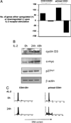
IL-2R signaling mediates a distinct pattern of gene transcription and cell cycle progression in CD4+CD25+ T cells. A, CD4+CD25+ T cells or rested primed CD4+ T cells were cultured in medium or medium plus IL-2 (100 U/ml) for 12 h. Total RNA was amplified and labeled with biotinylated ribonucleotides before hybridization to MGU74Av2 chips. Chips were subsequently stained and scanned as per manufacturers instructions. The number of genes with significantly increased or decreased expression (at least 2-fold) in two independent experiments per individual stimulation was determined using Microarray Suite (MAS 5.0) software. CD4+CD25+ T cells sorted by flow cytometry were either analyzed immediately or cultured in medium with IL-2 for the times indicated. B, Cells were lysed and probed with anti-cyclin D3, anti-p27kip, anti-myc, or anti-actin by Western blot as indicated. C, CD4+CD25+ cells and primed CD4+ T cells were stimulated with IL-2, fixed in cold ethanol, and stained for DNA content with propidium iodide. Results are representative of three independent experiments.
FIGURE 4
FIGURE 4
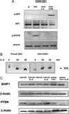
IL-2 activates PI3K and negative regulation of PI3K signaling occurs downstream of PI3K. A, Purified CD4+CD25+ T cells were stimulated with IL-2 (100 U/ml) in combination with latex beads precoated with mAbs to anti-CD3 (2 µg/ml) and anti-CD28 (10 µg/ml) as indicated for 30 min. Cell lysates (1 × 10⁶ cells/sample) were prepared and analyzed as indicated. B, Primed CD4+ T cells and purified CD4+CD25+ were stimulated with IL-2 (100 U/ml) for the times indicated. Cells were lysed, PI3K was immunoprecipitated overnight, and specific enzyme activity was measured by in vitro kinase reaction and TLC as per Materials and Methods. Migration of specific PIP3 product was determined using unlabelled PIP3 and revealed using iodine crystals. Results are representative of three experiments. C, Isolated CD4+CD25+ or CD4+CD25 T cells were either analyzed immediately or cultured in medium with 100 U/ml IL-2 for 96 h. Activated CD4+ T cells were rested overnight and stimulated with 100 U/ml IL-2 for 24 h. Samples were lysed and probed with anti-SHIP, anti-PTEN, or anti-actin by Western blot as indicated. Results are representative of three experiments.
FIGURE 5
FIGURE 5
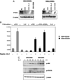
Activation of TCR down-regulates PTEN and restores IL-2-mediated PI3K signaling. A, Purified CD4+CD25+ and CD4+CD25 T cells were stimulated with anti-CD3 (0.5 µg/ml) alone or in combination with anti-CD28 (2 µg/ml) or IL-2 (100 U/ml) and T-depleted, irradiated APCs for 72 h. Samples were lysed and probed with anti-PTEN or anti-actin by Western blot as indicated. Results are representative of three experiments. B, CFSE labeled CD4+CD25+ and CD4+CD25 T cells were stimulated with anti-CD3 (0.5 µg/ml), anti-CD28 (5 µg/ml), IL-2 100 U/ml and T-depleted, irradiated APCs for 48 h and analyzed by flow cytometry (data not shown). Cells were subsequently washed and replated in complete medium alone or with IL-2 (100 U/ml) for 48 h. [3H]Thymidine was added to cultures for the last 16 h. C, Purified CD4+CD25+ and CD4+CD25 T cells were stimulated with anti-CD3 (0.5 µg/ml), anti-CD28 (5 µg/ml) and T-depleted, irradiated APCs for 72 h, then rested for 6 h in complete medium. Cells were subsequently stimulated with 100 U/ml IL-2 for 30 min. Samples were lysed and probed by Western blot as indicated.

References

    1. Waldmann TA, Dubois S, Tagaya Y. Contrasting roles of IL-2 and IL-15 in the life and death of lymphocytes: implications for immunotherapy. Immunity. 2001;14:105. - PubMed
    1. Refaeli Y, Van Parijs L, London CA, Tschopp J, Abbas AK. Biochemical mechanisms of IL-2-regulated Fas-mediated T cell apoptosis. Immunity. 1998;8:615. - PubMed
    1. Malek TR, Yu A, Vincek V, Scibelli P, Kong L. CD4 regulatory T cells prevent lethal autoimmunity in IL-2Rβ-deficient mice: implications for the nonredundant function of IL-2. Immunity. 2002;17:167. - PubMed
    1. Murakami M, Sakamoto A, Bender J, Kappler J, Marrack P. CD25+CD4+T cells contribute to the control of memory CD8+T cells. Proc. Natl. Acad. Sci. USA. 2002;99:8832. - PMC - PubMed
    1. Almeida AR, Legrand N, Papiernik M, Freitas AA. Homeostasis of peripheral CD4+T cells: IL-2Rα and IL-2 shape a population of regulatory cells that controls CD4+ T cell numbers. J. Immunol. 2002;169:4850. - PubMed

Publication types

MeSH terms