. 2004 May;16(5):1263-75.
doi: 10.1105/tpc.021030. Epub 2004 Apr 20.

Cotyledon vascular pattern2-mediated inositol (1,4,5) triphosphate signal transduction is essential for closed venation patterns of Arabidopsis foliar organs

Affiliations

Cotyledon vascular pattern2-mediated inositol (1,4,5) triphosphate signal transduction is essential for closed venation patterns of Arabidopsis foliar organs

Francine M Carland et al. Plant Cell. 2004 May.

Abstract

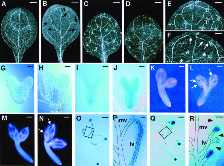
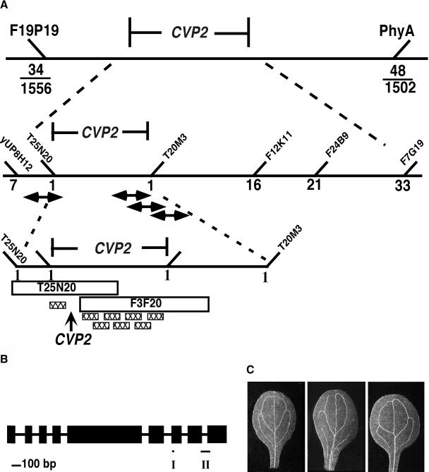
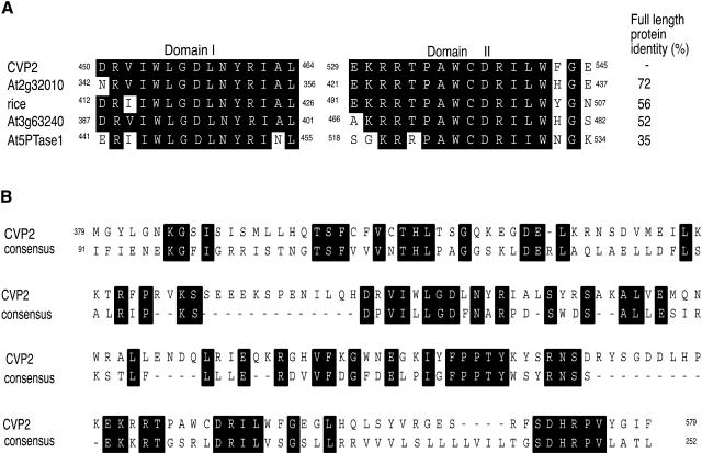
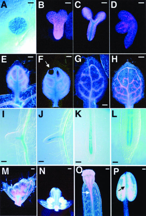
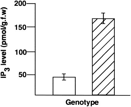
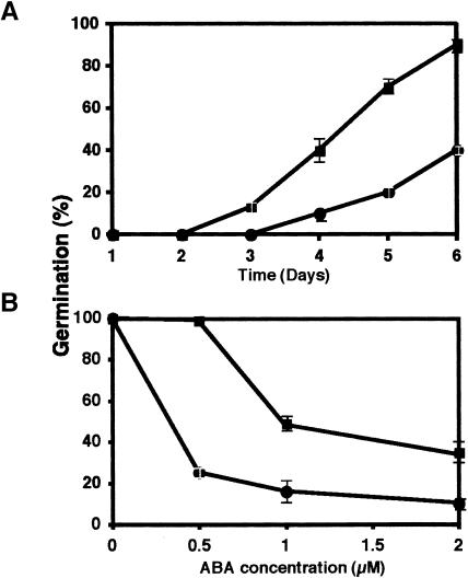
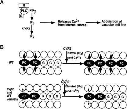
Vein patterns in leaves and cotyledons form in a spatially regulated manner through the progressive recruitment of ground cells into vascular cell fate. To gain insight into venation patterning mechanisms, we have characterized the cotyledon vascular pattern2 (cvp2) mutants, which exhibit an increase in free vein endings and a resulting open vein network. We cloned CVP2 by a map-based cloning strategy and found that it encodes an inositol polyphosphate 5' phosphatase (5PTase). 5PTases regulate inositol (1,4,5) triphosphate (IP(3)) signal transduction by hydrolyzing IP(3) and thus terminate IP(3) signaling. CVP2 gene expression is initially broad and then gradually restricted to incipient vascular cells in several developing organs. Consistent with the inferred enzymatic activity of CVP2, IP(3) levels are elevated in cvp2 mutants. In addition, cvp2 mutants exhibit hypersensitivity to the plant hormone abscisic acid. We propose that elevated IP(3) levels in cvp2 mutants reduce ground cell recruitment into vascular cell fate, resulting in premature vein termination and, thus, in an open reticulum.


Figures

Figure 1.
Figure 1.
CVP2 Acts Early in Procambial Patterning during Embryogenesis. (A) to (F) Cleared specimens viewed with dark-field optics. Wild-type cotyledon (A), cvp2 cotyledon (B), wild-type leaf (C), and cvp2 leaf (D) are shown. Higher magnification of (C) and (D) are shown in (E) and (F), respectively. With dark-field optics, trichomes (t) appear translucent with three spikes. (G) to (N) Athb8:GUS embryo expression. Wild-type heart (G), cvp2 heart (H), wild-type torpedo (I), cvp2 torpedo (J), wild-type walking stick (K), cvp2 walking stick (L), wild-type bent cotyledon (M), and cvp2 bent cotyledon (N) are shown. Dark-field images are more sensitive than Nomarski optical images and show weak GUS expression as pink and strong GUS expression as blue. (O) to (R) DR5:GUS expression of bent cotyledon stage embryo. Wild-type embryo (O), wild-type proximal lateral vein at a higher magnification of the boxed region in (O) to show uniform DR5:GUS expression (P), cvp2 embryo (Q), and cvp2 proximal lateral vein at a higher magnification of the boxed region in (Q) to show short stretches of DR5:GUS expression in cvp2 mutants (R). Arrowheads designate absence of DR5:GUS. In wild-type embryos, 369 out of 394 lateral veins showed continuous DR5:GUS expression. In cvp2 embryos, 33 out of 330 lateral veins showed continuous DR5:GUS expression. Roots of embryos in (O) and (Q) were distorted while separating cotyledons. Vein patterns are outlined in (P) and (R). Arrows indicate free vein endings of secondary veins; asterisks indicate free vein endings of tertiary veins. Dark-field optics were used for (A) to (F) and (K) to (N); Nomarski optics were used for (G) to (J) and (O) to (R). vi, vascular island; mv, midvein; lv, lateral vein. Bars = 500 μM in (A) to (D), 250 μM in (E) and (F), 10 μM in (G), (H), (P), and (R), 25 μM in (I) and (J), 50 μM in (K) and (L), and 80 μM in (M), (N), (O), and (Q).
Figure 2.
Figure 2.
Map-Based Cloning of CVP2. (A) Scheme showing the map-based cloning of CVP2. CVP2 was localized to the top of chromosome one, represented by a solid horizontal line, using the flanking markers PhyA and F19P19. For PhyA and F19P19, the fractions indicate the number of recombinants over the total number of meiotic events. The numbers of recombinants for additional markers are shown below the diagonal line. The name of the BAC clone (45° angle) is given for a specific polymorphic marker that was derived from the BAC. Double-headed arrows represent TACs that were identified in the region by hybridization studies. Hatched boxes represent some of the clones that were found to reside in the vicinity of CVP2 and assayed for cvp2 complementation. Because none of these clones rescued the cvp2 mutant phenotype, CVP2 was localized to one of four genes on BAC T25N20 that were not represented in the libraries. (B) CVP2 gene structure. Solid vertical bars represent exons. Exon/intron junctions were determined by comparing a CVP2 cDNA isolated from seedling tissue with the genomic sequence in the database. Mutations identified in four cvp2 alleles are given. Specifically, cvp2-1 has a G-to-A nucleotide substitution that substitutes a Gly residue for Asp at amino acid 567; cvp2-2 contains a C-to-T nucleotide substitution that changes an Ala to a Val in the PAWCDRIL (amino acid 535) site of domain II; cvp2-3 contains a G-to-A nucleotide substitution that changes a Trp to a stop codon at amino acid 144; cvp2-4 (diepoxybutane mutant) has an A-to-T nucleotide substitution at the fourth exon/intron junction (amino acid 136), which is predicted to cause a splicing error resulting in a misread message for 45 residues before termination. Short horizontal bars represent 5PTase signature motifs, domains I and II. (C) Complementation of cvp2 with 5PTase. A genomic clone of At1g05470 containing the coding region and 1.5 kb upstream of the translational start codon was introduced into cvp2-1 mutants. Cleared specimens were viewed with dark-field optics. Left, the wild type; center, cvp2-1; right, cvp2-1/At1g05470.
Figure 3.
Figure 3.
CVP2 Is a Member of the Endonuclease/Exonuclease/Phosphatase Family. (A) Alignment of CVP2 with other plant 5PTases. The accession number for the rice 5PTase is AA037964. (B) CVP2 is aligned to a consensus sequence for endonuclease/exonuclease/phosphatase family members according to the NCBI conserved domain database (http://www.ncbi.nlm.nih.gov/Structure/cdd). The C-terminal part of the CVP2 protein is shown. Members with accession numbers for which the consensus sequence was derived include the following: S. pombe synaptojanin, 1I9Y_A; leptospira hemolytic protein, AAB68647; S. pombe RNA nuclease, CAB42372; C. elegans reverse transcriptase, CAB07490; catabolite repressor protein, ACC44428; Homo sapiens Type I 5PTase, Q14642; bovine DNase I, 1DNK_A; Escherichia coli Exonuclease III, 1AKO; and human endonuclease I 1DEB_B. Alignments were conducted using MacVector 7.0 (Oxford Molecular, Madison, WI). Identical residues are shaded black.
Figure 4.
Figure 4.
CVP2 Expression by GUS Histochemical Staining. (A) to (D) Embryos at globular (A), torpedo (B), late torpedo (C), and walking-stick stages (D). (E) to (H) Sequential series of developing first rosette leaf. (E) Young leaf to show strong CVP2 expression in procambial cells and weaker expression in areoles. (F) Slightly more mature leaf to show the decrease in weak areole CVP2 expression at leaf apex (arrow) reflecting the basipetal maturation of the leaf. (G) More advanced leaf that demonstrates CVP2 expression in all developing veins. (H) CVP2 expression is restricted to developing veins in fully developed leaf. Note the absence of GUS staining in the apical loop in which vascular cells have already differentiated as tracheary elements and appear as white cells. (I) to (L) Root expression. (I) and (J) CVP2 is expressed in the procambial core of the emerging lateral root. (K) and (L) CVP2 expression is limited to root tip, specifically to two procambial cell files. (M) to (P) Floral expression of CVP2. CVP2 is broadly expressed in inflorescence (M) and then restricted to developing floral organ veins, such as those in the sepal (N), gynoecium (O), and anther (P). Dark-field optics were used for (B) to (H) and (M) to (P); Nomarski optics were used for (A) and (I) to (L). Bars = 10 μM in (A), 25 μM in (B), (I), and (L), 40 μM in (C), 50 μM in (D), (E), (F), (J), (K), and (P), 60 μM in (G), 100 μM in (H), (M), and (O), and 200 μM in (N).
Figure 5.
Figure 5.
IP3 Levels Are Elevated in cvp2 Mutants. Experiments were done in triplicate and error bars indicate standard error. Open, the wild type; hatched, cvp2. g.f.w., grams of fresh weight.
Figure 6.
Figure 6.
cvp2 Response to ABA. Seed germination was measured on increasing doses of ABA for 6 d. Wild-type and cvp2 seed germination on 2 μM ABA over a 6-d period (A) and on day 4 with increasing amounts of ABA (B). Squares, the wild type; circles, cvp2. Values represent triplicate experiments, and error bars show standard error.
Figure 7.
Figure 7.
Model for the Role of CVP2. (A) Hydrolyzing activity of CVP2. This model is based on animal systems in which the role of 5PTases is better understood. In response to as yet unidentified external stimulus, a membrane receptor activates phospholipase C (PLC)/G protein complexes (G) to breakdown PIP2 into IP3 (and diacylgylcerol). Acting as a second messenger, IP3 amplifies the primary signal by releasing Ca2+ from internal stores. The downstream biological response for the appropriate Ca2+ parameters is acquisition of vascular cell fate. CVP2 serves to hydrolyze IP3 into the inactive form, inositol (1,4) diphosphate (IP2), and thus terminates IP3 signal transduction. (B) Progessing procambial strand (PC) encounters ground cells (G). In the wild type, CVP2 would act to prevent sustained Ca2+ oscillations by hydrolyzing IP3 into IP2. In this manner, the correct Ca2+ signature is perceived by downstream Ca2+-dependent target genes, resulting in the recruitment of ground cells into vascular cell fate. In cvp2 foliar organs and in freely terminating wild-type veinlets, because of the absence of CVP2 hydrolytic activity, IP3 levels are elevated, resulting in sustained Ca2+ release. The inappropriate Ca2+ levels reduce ground cell specification into procambial cells, presumably by misregulated Ca2+ response genes, and the veins prematurely terminate. The decrease in size and number of arrows from the procambial cells indicates that the radial proliferation of cvp2 veins is also affected.

Similar articles

Cited by

References

    1. Allen, G.J., Chu, S.P., Harrington, C.L., Schumacher, K., Hoffmann, T., Tang, Y.Y., Grill, E., and Schroeder, J.I. (2001). A defined range of guard cell calcium oscillation parameters encodes stomatal movements. Nature 411, 1053–1057. - PubMed
    1. Baima, S., Nobili, F., Sessa, G., Lucchetti, S., Ruberti, I., and Morelli, G. (1995). The expression of the Athb-8 homeobox gene is restricted to provascular cells in Arabidopsis thaliana. Development 121, 4171–4182. - PubMed
    1. Berdy, S.E., Kudla, J., Gruissem, W., and Gillaspy, G.E. (2001). Molecular characterization of At5PTase1, an inositol phosphatase capable of terminating inositol trisphosphate signaling. Plant Physiol. 126, 801–810. - PMC - PubMed
    1. Berleth, T., and Jurgens, G. (1993). The role of the monopteros gene in organizing the basal body region of the Arabidopsis embryo. Development 118, 575–587.
    1. Berridge, M.J. (1993). Inositol trisphosphate and calcium signalling. Nature 361, 315–325. - PubMed

MeSH terms

Substances

Associated data