

Structural variant of the intergenic internal ribosome entry site elements in dicistroviruses and computational search for their counterparts
- PMID: 15100433
- PMCID: PMC1370568
- DOI: 10.1261/rna.5208104
Structural variant of the intergenic internal ribosome entry site elements in dicistroviruses and computational search for their counterparts
Abstract
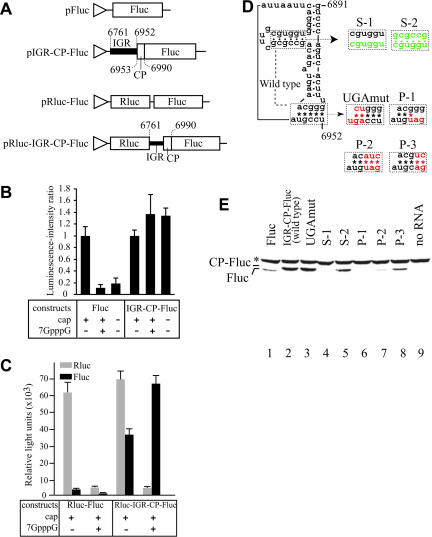
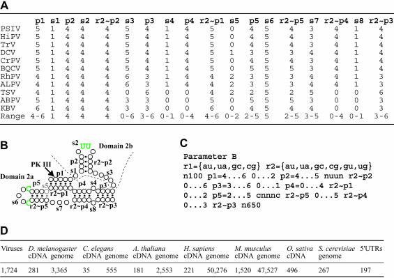
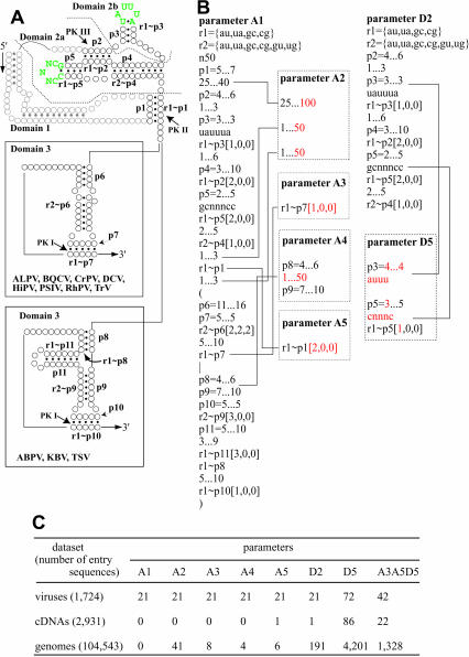
The intergenic region (IGR) located upstream of the capsid protein gene in dicistroviruses contains an internal ribosome entry site (IRES). Translation initiation mediated by the IRES does not require initiator methionine tRNA. Comparison of the IGRs among dicistroviruses suggested that Taura syndrome virus (TSV) and acute bee paralysis virus have an extra side stem loop in the predicted IRES. We examined whether the side stem is responsible for translation activity mediated by the IGR using constructs with compensatory mutations. In vitro translation analysis showed that TSV has an IGR-IRES that is structurally distinct from those previously described. Because IGR-IRES elements determine the translation initiation site by virtue of their own tertiary structure formation, the discovery of this initiation mechanism suggests the possibility that eukaryotic mRNAs might have more extensive coding regions than previously predicted. To test this hypothesis, we searched full-length cDNA databases and whole genome sequences of eukaryotes using the pattern matching program, Scan For Matches, with parameters that can extract sequences containing secondary structure elements resembling those of IGR-IRES. Our search yielded several sequences, but their predicted secondary structures were suggested to be unstable in comparison to those of dicistroviruses. These results suggest that RNAs structurally similar to dicistroviruses are not common. If some eukaryotic mRNAs are translated independently of an initiator methionine tRNA, their structures are likely to be significantly distinct from those of dicistroviruses.
Figures
Similar articles
-
Factor-independent assembly of elongation-competent ribosomes by an internal ribosome entry site located in an RNA virus that infects penaeid shrimp.J Virol. 2005 Jan;79(2):677-83. doi: 10.1128/JVI.79.2.677-683.2005. J Virol. 2005. PMID: 15613295 Free PMC article.
-
Functional Insights into the Adjacent Stem-Loop in Honey Bee Dicistroviruses That Promotes Internal Ribosome Entry Site-Mediated Translation and Viral Infection.J Virol. 2018 Jan 2;92(2):e01725-17. doi: 10.1128/JVI.01725-17. J Virol. 2018. PMID: 29093099 Free PMC article.
-
The Hinge Region of the Israeli Acute Paralysis Virus Internal Ribosome Entry Site Directs Ribosomal Positioning, Translational Activity, and Virus Infection.J Virol. 2022 Mar 9;96(5):e0133021. doi: 10.1128/JVI.01330-21. Epub 2022 Jan 12. J Virol. 2022. PMID: 35019716 Free PMC article.
-
Functional analysis of structural motifs in dicistroviruses.Virus Res. 2009 Feb;139(2):137-47. doi: 10.1016/j.virusres.2008.06.006. Epub 2008 Jul 25. Virus Res. 2009. PMID: 18621089 Review.
-
Comparing the three-dimensional structures of Dicistroviridae IGR IRES RNAs with other viral RNA structures.Virus Res. 2009 Feb;139(2):148-56. doi: 10.1016/j.virusres.2008.07.007. Epub 2008 Aug 15. Virus Res. 2009. PMID: 18672012 Free PMC article. Review.
Cited by
-
A distinct group of hepacivirus/pestivirus-like internal ribosomal entry sites in members of diverse picornavirus genera: evidence for modular exchange of functional noncoding RNA elements by recombination.J Virol. 2007 Jun;81(11):5850-63. doi: 10.1128/JVI.02403-06. Epub 2007 Mar 28. J Virol. 2007. PMID: 17392358 Free PMC article.
-
IRESpy: an XGBoost model for prediction of internal ribosome entry sites.BMC Bioinformatics. 2019 Jul 30;20(1):409. doi: 10.1186/s12859-019-2999-7. BMC Bioinformatics. 2019. PMID: 31362694 Free PMC article.
-
Factor-independent assembly of elongation-competent ribosomes by an internal ribosome entry site located in an RNA virus that infects penaeid shrimp.J Virol. 2005 Jan;79(2):677-83. doi: 10.1128/JVI.79.2.677-683.2005. J Virol. 2005. PMID: 15613295 Free PMC article.
-
Structural determinants of an internal ribosome entry site that direct translational reading frame selection.Nucleic Acids Res. 2014 Aug;42(14):9366-82. doi: 10.1093/nar/gku622. Epub 2014 Jul 18. Nucleic Acids Res. 2014. PMID: 25038250 Free PMC article.
-
Viral IRES RNA structures and ribosome interactions.Trends Biochem Sci. 2008 Jun;33(6):274-83. doi: 10.1016/j.tibs.2008.04.007. Epub 2008 May 28. Trends Biochem Sci. 2008. PMID: 18468443 Free PMC article. Review.
References
Publication types
MeSH terms
Substances
LinkOut - more resources
Full Text Sources