. 2004 May;52(3):801-14.
doi: 10.1111/j.1365-2958.2004.04016.x.

EzrA prevents aberrant cell division by modulating assembly of the cytoskeletal protein FtsZ

Affiliations

EzrA prevents aberrant cell division by modulating assembly of the cytoskeletal protein FtsZ

Daniel P Haeusser et al. Mol Microbiol. 2004 May.

Abstract

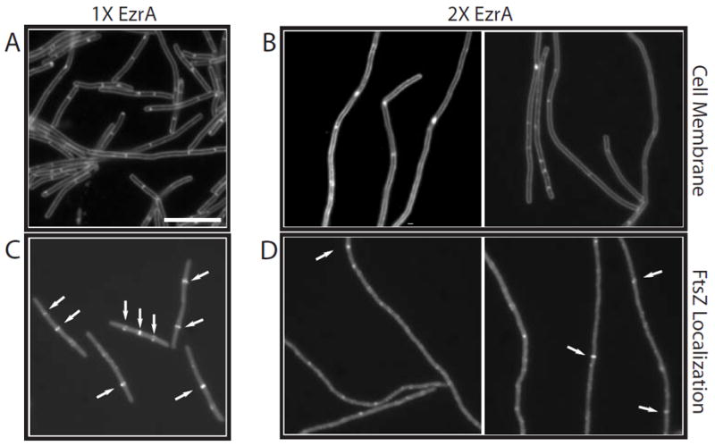
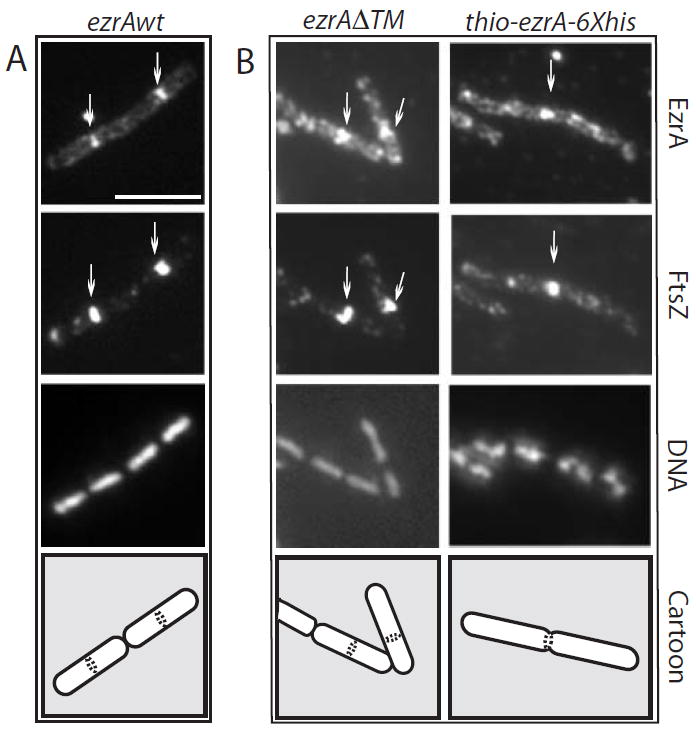
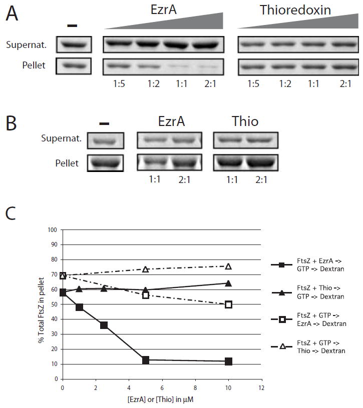
In response to a cell cycle signal, the cytoskeletal protein FtsZ assembles into a ring structure that establishes the location of the division site and serves as a framework for assembly of the division machinery. A battery of factors control FtsZ assembly to ensure that the ring forms in the correct position and at the precise time. EzrA, a negative regulator of FtsZ ring formation, is important for ensuring that the ring forms only once per cell cycle and that cytokinesis is restricted to mid-cell. EzrA is distributed throughout the plasma membrane and localizes to the ring in an FtsZ-dependent manner, suggesting that it interacts directly with FtsZ to modulate assembly. We have performed a series of experiments examining the interaction between EzrA and FtsZ. As little as twofold overexpression of EzrA blocks FtsZ ring formation in a sensitized genetic background, consistent with its predicted function. A purified EzrA fusion protein interacts directly with FtsZ to block assembly in vitro. Although EzrA is able to inhibit FtsZ assembly, it is unable to disassemble preformed polymers. These data support a model in which EzrA interacts directly with FtsZ at the plasma membrane to prevent polymerization and aberrant FtsZ ring formation.


Figures

Fig. 1
Fig. 1
The effect of EzrA overexpression on cell division and Z-ring formation in ftsZts mutant cells. A and B. Cells were stained with the vital membrane stain FM4-64 to visualize septa. C and D. FtsZts-GFP. A and C. MO54 (amyE∷Pspachy-ezrA∷ftsZts) cells grown in the absence of inducer. B and D. MO54 cells 3 h after the addition of IPTG. EzrA levels are twofold above wild type. Arrows point to FtsZ rings. Scale bar = 5 μm.
Fig. 2
Fig. 2
The transmembrane anchor is dispensable for EzrA localization. Cells were grown to mid-exponential phase (OD600 = ≈ 0.5) in the presence of IPTG and fixed and stained for immunofluorescence microscopy using antiserum against FtsZ (Levin and Losick, 1996) or EzrA (this work). A single field of view is shown in each column. From top to bottom: EzrA, FtsZ, DNA and a schematic showing cell boundaries and the position of the EzrA/FtsZ rings. A. ezrA∷Pspachy-ezrA (PL923). B. ezrA∷Pspachy-ezrAΔTM (PL925). The high background in the anti-EzrA panels in (B) results from cytoplasmic staining of EzrA in the absence of the transmembrane domain. Scale bar = 5 μM.
Fig. 3
Fig. 3
Electron micrographs (EMs) of B. subtilis FtsZ assembled under different conditions. A. ‘Deep-etch’ EM of FtsZ polymers assembled in the presence of GTP. Micrograph kindly provided by Dr John Heuser. B–D. Negative stain EM of (B) and (C) FtsZ assembled in the presence of GTP and (D) FtsZ assembled in the presence of GTP and 0.1 mg ml−1 DEAE-dextran. Note the single-stranded polymers, paired protofilaments and small bundles of FtsZ in (B), hoop-shaped bundle in (C) and large bundles of filaments in (D). FtsZ was at 5 μM for both the ‘deep etch’ and the negative stain EM. GTP was at 1 mM for all experiments. The scale bar is 50 nm in (A) and 20 nm in (B) to (D).
Fig. 4
Fig. 4
EzrA inhibits sedimentation of FtsZ assembled in vitro. FtsZ is 5 μM in all reactions. The samples were spun at 250 000 g to separate unassembled protein from protofilament bundles, and the relative concentration of protein in the supernatants (unassembled FtsZ) and pellets (assembled FtsZ) was analysed by SDS–PAGE. A. EzrA and thioredoxin were added to final concentrations of 1, 2.5 μM, 5 μM and 10 μM before the addition of GTP and DEAE-dextran. Left. Increasing concentration of FtsZ in the supernatants and decreasing concentration of FtsZ in the pellets result from the inhibition of FtsZ assembly by purified EzrA. Right. The thioredoxin control protein does not significantly inhibit FtsZ assembly. The ratio of EzrA or thioredoxin to FtsZ is shown under each lane. B. Order of addition. EzrA and thioredoxin were added to the reaction 1 min after the addition of GTP. Ratios of EzrA and thioredoxin to FtsZ are shown below each lane. C. Plot of percentage of total FtsZ in the pellet. EzrA, squares; thioredoxin, triangles. Closed symbols represent data from the experiment shown in (A). Open symbols represent data from the experiment shown in (B). Differences in the sedimentation efficiency of FtsZ in the absence of EzrA or thioredoxin between the experiments shown in (A) and (B) reflect normal variations between protein preparations.
Fig. 5
Fig. 5
Quantification of EzrA protein in B. subtilis. Wild-type cells (JH642) grown to mid-exponential phase (OD600 = 0.5) in Luria broth (LB) were sampled for colony-forming units and lysed as described in Experimental procedures. Different amounts of lysate (6.29 × 10³ cfu μl−1) were loaded in lane 1 (5 μl) and lane 2 (10 μl). EzrA was detected by immunoblotting with whole sera. EzrA was quantified by comparing the intensity of the bands in lanes 1 and 2 with that of a twofold serial dilution of purified EzrA fusion protein (four right hand lanes) taking into account the number of colony-forming units. Based on these data, we estimate that there are 10 000–20 000 copies of EzrA per cell during exponential growth in LB.
Fig. 6
Fig. 6
EzrA inhibits FtsZ polymerization as measured by 90° angle light scattering. Light scattering is in arbitrary units (AU). FtsZ is at 2.5 μM in all reactions. A. EzrA or thioredoxin was added to the reaction before the addition of GTP to a final concentration of 5 μM. Arrow indicates the time of addition of 1 mM GTP or 1 mM GDP. The addition of GDP reduces FtsZ light scattering below baseline, presumably because it inhibits GTP-independent association of FtsZ monomers. B. Concentration-dependent inhibition of FtsZ assembly by EzrA. The ratio of EzrA to FtsZ is shown underneath each bar. The ratio of thioredoxin to FtsZ is 2:1. Margins of error were calculated using data from three independent experiments. C. EzrA or GDP were added to an FtsZ assembly reaction 1 min after the addition of GTP. The first arrow indicates the addition of GTP and the second the addition of EzrA or GDP. EzrA was at a final concentration of 5 μM in this experiment. GDP was used at a final concentration of 1 mM.
Fig. 7
Fig. 7
EzrA binds to FtsZ. A and B. Immunoblots of cross-linking reaction products. A. Anti-FtsZ. B. Anti-6×His. FtsZ alone (lane 1); EzrA alone (lane 2); FtsZ plus EzrA-6×His (lane 3); FtsZ plus Thio-6×His (lane 4); FtsZ plus Spo0J-6×His (lane 5). C and D. Co2+ affinity purification of 6×His-tagged cross-linked products. Eluants were separated by SDS–PAGE and subjected to immunoblotting with antisera against FtsZ (C) or 6×His (D). FtsZ plus EzrA-6×His (lane 1); Thio-6×His (lane 2); or Spo0J-6×His (lane 3). Breakdown products of EzrA-6×His can be seen in (D), lane 1. Molecular weight markers in kDa are shown to the right of blots (A) and (C). Variations in band intensity reflect differences in transfer efficiency between high- and low-molecular-weight material.
Fig. 8
Fig. 8
Model for EzrA inhibition of FtsZ assembly at the cell membrane. Left. EzrA functions at the membrane to inhibit FtsZ assembly and prevent aberrant Z-ring formation. At mid-cell, a positive acting factor (arrows) promotes FtsZ assembly and overcomes EzrA activity allowing for medial ring formation. Right. In the absence of EzrA, FtsZ is free to assemble anywhere along the inner surface of the membrane leading to aberrant FtsZ assembly at cell poles. Despite the formation of polar Z rings, a positive acting factor (arrows) still promotes FtsZ assembly at mid-cell and leads to a situation in which this position is favoured for cytokinesis.

Similar articles

Cited by

References

    1. Addinall SG, Lutkenhaus J. FtsZ-spirals and –arcs determine the shape of the invaginating septa in some mutants of Escherichia coli. Mol Microbiol. 1996;22:231–237. - PubMed
    1. Arakawa T, Timasheff SN. Mechanism of protein salting in and salting out by divalent cation salts: balance between hydration and salt binding. Biochemistry. 1984;23:5912–5923. - PubMed
    1. Beall B, Lutkenhaus J. Nucleotide sequence and insertional inactivation of a Bacillus subtilis gene that affects cell division, sporulation, and temperature sensitivity. J Bacteriol. 1989;171:6821–6834. - PMC - PubMed
    1. Beall B, Lutkenhaus J. Impaired cell division and sporulation of a Bacillus subtilis strain with the ftsA gene deleted. J Bacteriol. 1992;174:2398–2403. - PMC - PubMed
    1. de Boer PAJ, Crossley RE, Rothfield LI. Roles of MinC and MinD in the site-specific septation block mediated by the MinCDE system of Escherichia coli. J Bacteriol. 1992;174:63–70. - PMC - PubMed

Publication types

MeSH terms

LinkOut - more resources