Review
. 2004 Feb;14(2):86-93.
doi: 10.1016/j.tcb.2003.12.003.

Matrix remodeling during endochondral ossification

Affiliations
Review

Matrix remodeling during endochondral ossification

Nathalie Ortega et al. Trends Cell Biol. 2004 Feb.

Abstract

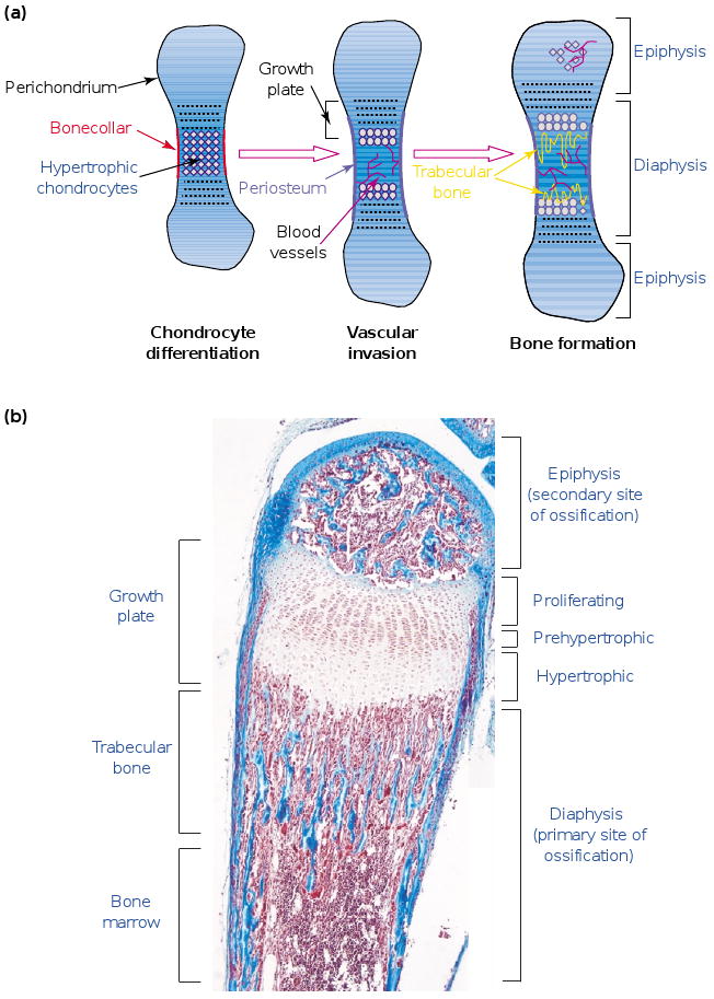
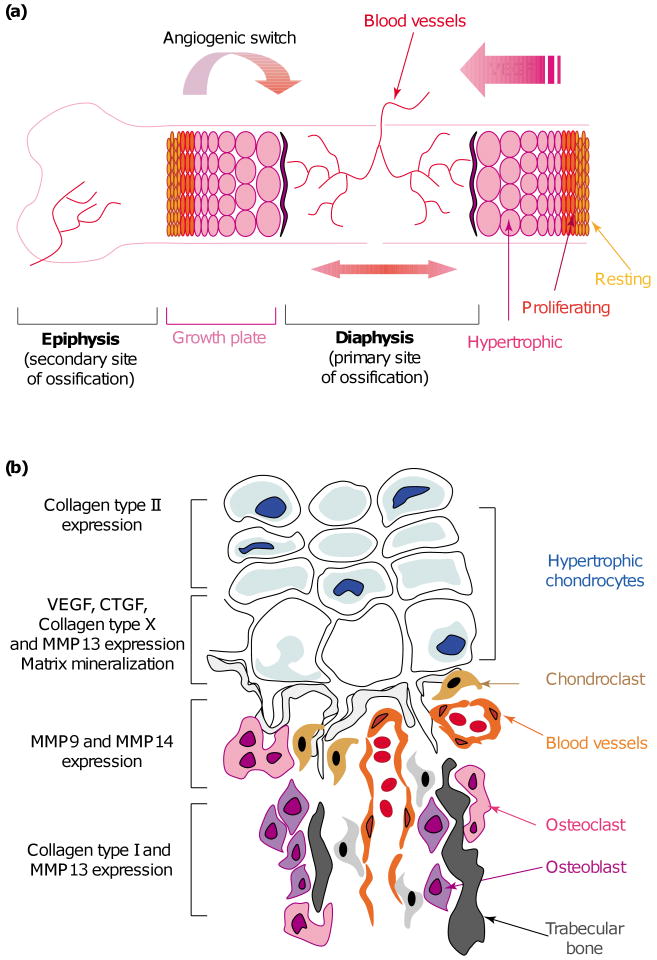
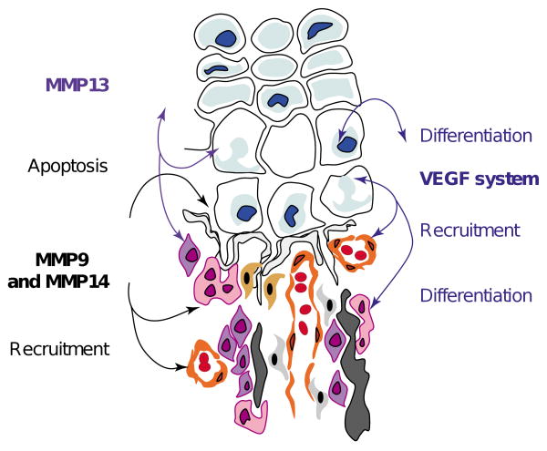
Endochondral ossification, the process by which most of the skeleton is formed, is a powerful system for studying various aspects of the biological response to degraded extracellular matrix (ECM). In addition, the dependence of endochondral ossification upon neovascularization and continuous ECM remodeling provides a good model for studying the role of the matrix metalloproteases (MMPs) not only as simple effectors of ECM degradation but also as regulators of active signal-inducers for the initiation of endochondral ossification. The daunting task of elucidating their specific role during endochondral ossification has been facilitated by the development of mice deficient for various members of this family. Here, we discuss the ECM and its remodeling as one level of molecular regulation for the process of endochondral ossification, with special attention to the MMPs.


Figures

Figure 1
Figure 1
The process of endochondral ossification. (a) During endochondral ossification, mesenchymal cells differentiate into chondrocytes and lead to the formation of cartilage templates. Vascularization occurs around these templates, and osteoblasts differentiate around the central area in the bone collar. Chondrocytes in this central area differentiate into hypertrophic chondrocytes and allow vascular invasion. After complete differentiation, they die and extracellular matrix (ECM) remodeling is carried out by osteoclasts and chondroclasts recruited together with the blood vessels. This remodeling is necessary for trabecular-bone synthesis by osteoblasts precursors and for the formation of the bone-marrow cavity. (b) A section of a two-week-old mouse metatarsal stained with Masson trichrome. Chondrocytes proliferate and differentiate into prehypertrophic and hypertrophic chondrocytes. Endochondral ossification first takes place at the primary site at the center of the diaphysis, which allows formation of the two growth plates. The growth plates are ultimately responsible for the elongation of the long bones. Later, endochondral ossification occurs at a secondary site, in the epiphysis of the long bones. Scale bar represents 200 μm.
Figure 2
Figure 2
Endochondral ossification, extracellular matrix (ECM) remodeling and angiogenic switch. (a) The process of endochondral ossification is dependent upon neovascularization. During this process, cartilage is replaced by bone. The differentiation of chondrocytes into hypertrophic chondrocytes is accompanied by the expression of angiogenic growth factors, notably VEGF, allowing vascular invasion of the cartilage. In the diaphysis, this invasion is strictly limited to the front of ossification in the last row of hypertrophic chondrocytes in direct contact with blood vessels. (b) The process of endochondral ossification also depends upon a switch in the expression of ECM molecules. Proliferating chondrocytes express collagen type II, whereas hypertrophic chondrocytes express collagen type X, matrix metalloprotease 13 (MMP13) and high levels of angiogenic growth factors VEGF and CTGF. On the other side of the ossification front, osteoclasts express MMP9, whereas osteoblasts express MMP13 and collagen type I.
Figure 3
Figure 3
Multiple roles of matrix metalloproteases (MMPs) in endochondral ossification. MMPs are directly involved in the remodeling of the extracellular matrix (ECM) through degradation of proteins such as collagens or proteoglycans. In addition, they have been implicated in more subtle functions such as apoptosis and the recruitment of cells, through the regulation of bioavailability and/or activation of factors sequestered into the ECM. For example, VEGF availability is regulated by this type of sequestration and release.

Similar articles

Cited by

References

    1. Karsenty G, Wagner EF. Reaching a genetic and molecular understanding of skeletal development. Dev Cell. 2002;2:389–406. - PubMed
    1. Costell M, et al. Perlecan maintains the integrity of cartilage and some basement membranes. J Cell Biol. 1999;147:1109–1122. - PMC - PubMed
    1. Arikawa-Hirasawa E, et al. Perlecan is essential for cartilage and cephalic development. Nat Genet. 1999;23:354–358. - PubMed
    1. Jacenko O, et al. A dominant interference collagen X mutation disrupts hypertrophic chondrocyte pericellular matrix and glycosaminoglycan and proteoglycan distribution in transgenic mice. Am J Pathol. 2001;159:2257–2269. - PMC - PubMed
    1. Blair HC, et al. Mechanisms balancing skeletal matrix synthesis and degradation. Biochem J. 2002;364:329–341. - PMC - PubMed

Publication types

MeSH terms

Substances

LinkOut - more resources