

doi: 10.1136/adc.2003.027334.
The magnetic resonance revolution in brain imaging: impact on neonatal intensive care
Affiliations
- PMID: 15102717
- PMCID: PMC1721681
- DOI: 10.1136/adc.2003.027334
Item in Clipboard
The magnetic resonance revolution in brain imaging: impact on neonatal intensive care
Arch Dis Child Fetal Neonatal Ed.
2004 May.
No abstract available
Figures
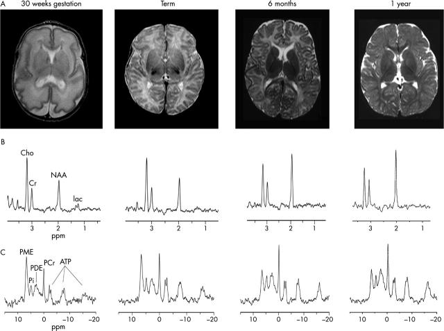
(A) Representative T2 weighted magnetic resonance (MR) images, (B) 1H MR spectra, and (C) 31P MR spectra from healthy infants at 30 weeks gestation, term, 6 months, and 1 year of age. (A) The MR images show an increase in volume, surface area, and sulcation of cerebral cortex and in volume and microstructural organisation of cerebral white matter with development. (B) The series of 1H MR spectra show a steady increase in N-acetyl aspartate (NAA; a marker of neuronal and axonal density and viability) and a decrease in brain lactate with maturation. (C) The series of 31P MR spectra show changing ratios of brain phospholipids and increasing energy state with maturation. PME, Phosphomonoesters; Pi, inorganic phosphate; PDE, phosphodiesters; PCr, phosphocreatine; ATP, adenosine triphosphate; Cho, choline; Cr, creatine; lac, lactate.
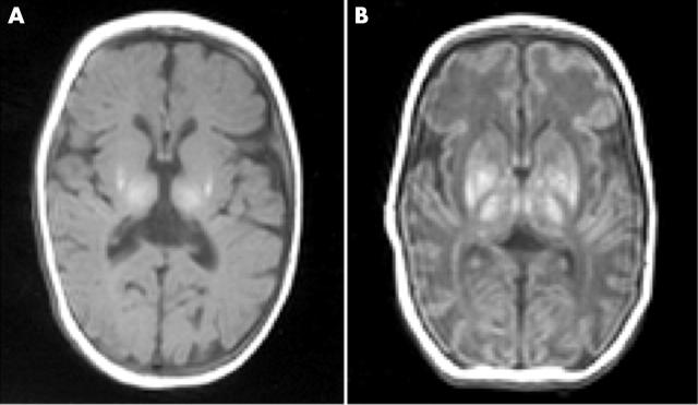
T1 weighted magnetic resonance images of (A) focal injury in an infant with Sarnat stage II neonatal encephalopathy (NE) aged 11 days and (B) global injury in an infant with Sarnat stage III NE aged 12 days. (A) Focal high signal intensity lesions are visible in the lentiform nuclei and thalami, and there is loss of the normal high signal intensity from myelin in the posterior limb of the internal capsule. (B) There are extensive high signal intensity lesions in the lentiform nuclei and thalami, loss of the normal high signal intensity from myelin in the posterior limb of the internal capsule, and abnormal low signal intensity in the white matter with loss of the normal grey/white matter differentiation.
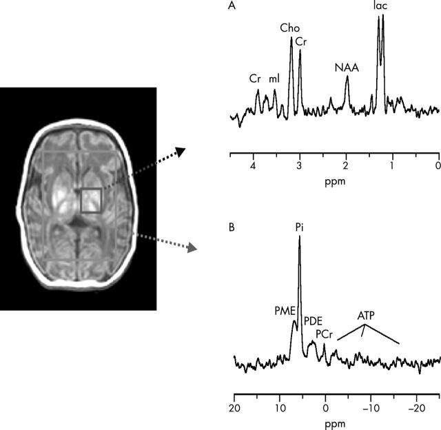
(A) 1H MR spectrum from left basal ganglia showing appreciably raised lactate and reduced N-acetyl aspartate (NAA). (B) 31P MR spectrum from the whole brain showing severe secondary energy failure with phosphocreatine (PCr) and ATP depletion and increased inorganic phosphate (Pi). The brain pHi was alkaline (7.18). PME, Phosphomonoesters; PDE, phosphodiesters; Cho, choline; Cr, creatine; mI, myo-inositol; lac, lactate.
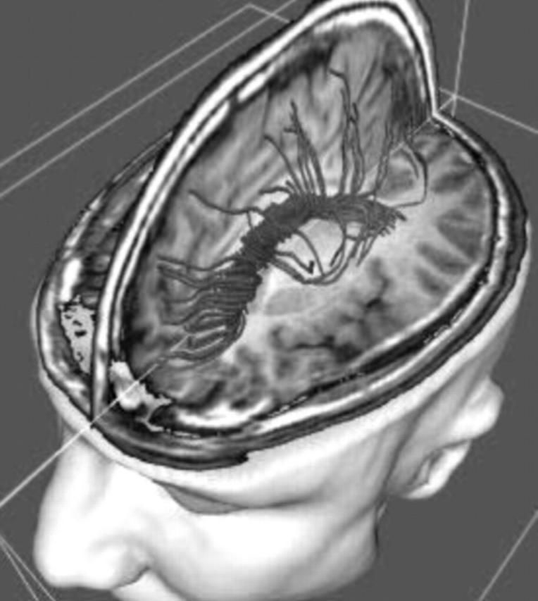
Fibre tracking which in the future may be possible using diffusion tensor imaging. If successful, this technique has huge potential in furthering our understanding of preterm white matter damage.
Similar articles
-
[Cerebral brain injury in preterm infants and the role of neuroimaging in predicting neurodevelopmental outcomes].Rev Med Suisse. 2014 Feb 19;10(418):442-9. Rev Med Suisse. 2014. PMID: 24640280 French.
-
The predictive validity of neonatal MRI for neurodevelopmental outcome in very preterm children.Semin Perinatol. 2015 Mar;39(2):147-58. doi: 10.1053/j.semperi.2015.01.008. Epub 2015 Feb 25. Semin Perinatol. 2015. PMID: 25724792 Review.
-
[Epoch-making ideas in care of skull injuries. New intensive care based on integrated physiology].Lakartidningen. 1994 Dec 14;91(50):4729-37. Lakartidningen. 1994. PMID: 7830424 Review. Swedish. No abstract available.
-
Advances in magnetic resonance imaging of the injured neonatal brain.Pediatr Ann. 2008 Jun;37(6):395-402. doi: 10.3928/00904481-20080601-09. Pediatr Ann. 2008. PMID: 18616193 Review. No abstract available.
-
Magnetic resonance imaging of the neonatal brain.Hosp Med. 1998 Jan;59(1):41-5. Hosp Med. 1998. PMID: 9798564 Review.
Cited by
-
Role of cerebral function monitoring in the newborn.Arch Dis Child Fetal Neonatal Ed. 2005 May;90(3):F201-7. doi: 10.1136/adc.2004.062745. Arch Dis Child Fetal Neonatal Ed. 2005. PMID: 15846008 Free PMC article. Review.
-
Fractional anisotropy in white matter tracts of very-low-birth-weight infants.Pediatr Radiol. 2007 Dec;37(12):1216-23. doi: 10.1007/s00247-007-0626-7. Epub 2007 Oct 2. Pediatr Radiol. 2007. PMID: 17909782 Free PMC article.
-
Post mortem magnetic resonance imaging in the fetus, infant and child: a comparative study with conventional autopsy (MaRIAS Protocol).BMC Pediatr. 2011 Dec 22;11:120. doi: 10.1186/1471-2431-11-120. BMC Pediatr. 2011. PMID: 22192497 Free PMC article.
-
Pilot randomized trial of therapeutic hypothermia with serial cranial ultrasound and 18-22 month follow-up for neonatal encephalopathy in a low resource hospital setting in Uganda: study protocol.Trials. 2011 Jun 4;12:138. doi: 10.1186/1745-6215-12-138. Trials. 2011. PMID: 21639927 Free PMC article. Clinical Trial.
-
Neonatal cranial ultrasound versus MRI and neurodevelopmental outcome at school age in children born preterm.Arch Dis Child Fetal Neonatal Ed. 2005 Nov;90(6):F489-93. doi: 10.1136/adc.2005.073908. Epub 2005 Jun 14. Arch Dis Child Fetal Neonatal Ed. 2005. PMID: 15956095 Free PMC article.
References
MeSH terms
LinkOut - more resources
Full Text Sources
Medical