

Alcohol exacerbates murine pulmonary tuberculosis
- PMID: 15102763
- PMCID: PMC387844
- DOI: 10.1128/IAI.72.5.2556-2563.2004
Alcohol exacerbates murine pulmonary tuberculosis
Abstract
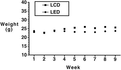
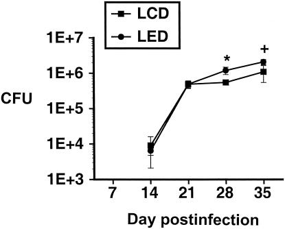
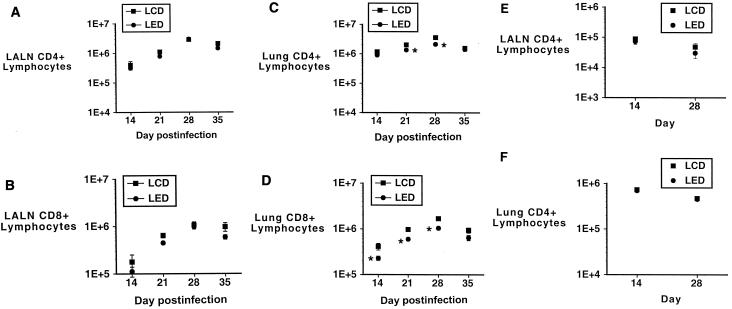
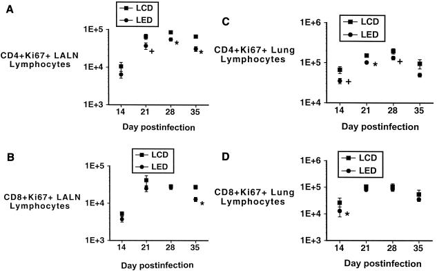
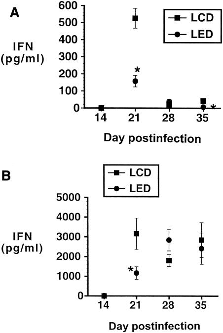
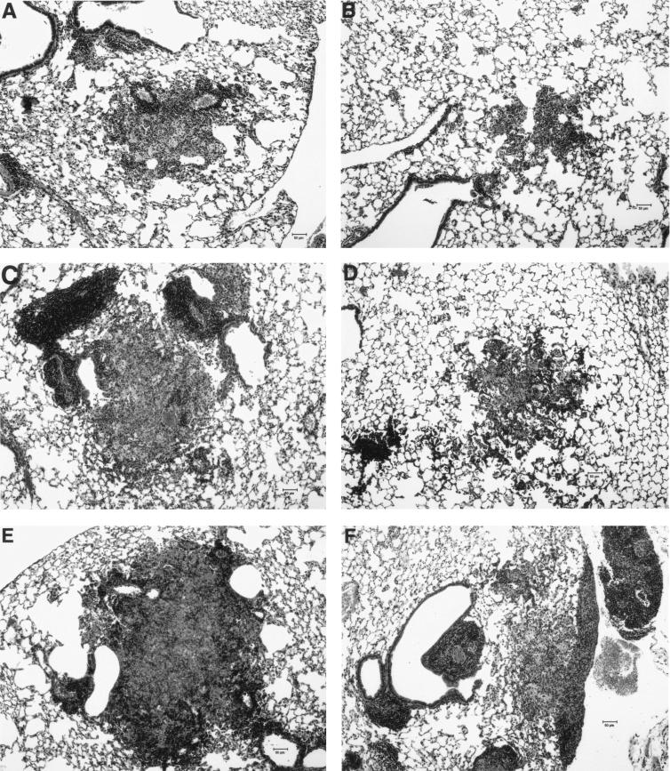
Alcohol consumption has been described as a risk factor for infection with Mycobacterium tuberculosis, but its contribution to tuberculosis has been difficult to isolate from other adverse socioeconomic factors. Our objective was to evaluate the impact of alcohol consumption on pulmonary infection with M. tuberculosis in a murine model. BALB/c mice were maintained on the Lieber-DeCarli liquid ethanol diet or a liquid control diet and infected intratracheally with low-dose M. tuberculosis H37Rv. Lung organism burdens, lung and lung-associated lymph node CD4(+)- and CD8(+)- lymphocyte numbers and rates of proliferation, and CD4(+)-lymphocyte cytokine production levels were compared between the groups. The alcohol-consuming mice had significantly higher lung organism burdens than the control mice, and the CD4(+)- and CD8(+)-lymphocyte responses to pulmonary infection with M. tuberculosis were blunted in the alcohol group. Lymphocyte proliferation and production of gamma interferon were decreased in the CD4(+) lymphocytes from the alcohol-consuming mice. Additionally, lung granulomas were significantly smaller in the alcohol-consuming mice. In conclusion, murine alcohol consumption is associated with decreased control of pulmonary infection with M. tuberculosis, which is accompanied by alterations in the region-specific CD4(+)- and CD8(+)-lymphocyte responses and defective lung granuloma formation.
Figures
Similar articles
-
The impact of alcohol on BCG-induced immunity against Mycobacterium tuberculosis.Alcohol Clin Exp Res. 2012 Feb;36(2):310-7. doi: 10.1111/j.1530-0277.2011.01624.x. Epub 2011 Oct 20. Alcohol Clin Exp Res. 2012. PMID: 22014229 Free PMC article.
-
Induction of Mycobacterium tuberculosis-specific primary and secondary T-cell responses in interleukin-15-deficient mice.Infect Immun. 2005 May;73(5):2910-22. doi: 10.1128/IAI.73.5.2910-2922.2005. Infect Immun. 2005. PMID: 15845497 Free PMC article.
-
CD11c(+) CD103(+) cells of Mycobacterium tuberculosis-infected C57BL/6 but not of BALB/c mice induce a high frequency of interferon-γ- or interleukin-17-producing CD4(+) cells.Immunology. 2015 Apr;144(4):574-86. doi: 10.1111/imm.12411. Immunology. 2015. PMID: 25322675 Free PMC article.
-
Regulation of the human immune response in tuberculosis.Infect Agents Dis. 1996 Mar;5(2):82-97. Infect Agents Dis. 1996. PMID: 8721045 Review. No abstract available.
-
[Research progress of pulmonary tuberculosis local immune].Zhonghua Jie He He Hu Xi Za Zhi. 2014 Feb;37(2):119-21. Zhonghua Jie He He Hu Xi Za Zhi. 2014. PMID: 24796594 Review. Chinese. No abstract available.
Cited by
-
The prevalence of concurrent pulmonary and extrapulmonary tuberculosis in Uganda: a retrospective study.Ther Adv Infect Dis. 2022 Jun 30;9:20499361221107304. doi: 10.1177/20499361221107304. eCollection 2022 Jan-Dec. Ther Adv Infect Dis. 2022. PMID: 35795170 Free PMC article.
-
Tobacco smoking, alcohol drinking, diabetes, low body mass index and the risk of self-reported symptoms of active tuberculosis: individual participant data (IPD) meta-analyses of 72,684 individuals in 14 high tuberculosis burden countries.PLoS One. 2014 May 2;9(5):e96433. doi: 10.1371/journal.pone.0096433. eCollection 2014. PLoS One. 2014. PMID: 24789311 Free PMC article.
-
Impairment of Hematopoietic Precursor Cell Activation during the Granulopoietic Response to Bacteremia in Mice with Chronic-Plus-Binge Alcohol Administration.Infect Immun. 2017 Oct 18;85(11):e00369-17. doi: 10.1128/IAI.00369-17. Print 2017 Nov. Infect Immun. 2017. PMID: 28784931 Free PMC article.
-
Tea Drinking and Its Association with Active Tuberculosis Incidence among Middle-Aged and Elderly Adults: The Singapore Chinese Health Study.Nutrients. 2017 May 25;9(6):544. doi: 10.3390/nu9060544. Nutrients. 2017. PMID: 28587081 Free PMC article.
-
The American Association of Tissue Banks tissue donor screening for Mycobacterium tuberculosis-Recommended criteria and literature review.Transpl Infect Dis. 2024 Nov;26 Suppl 1(Suppl 1):e14294. doi: 10.1111/tid.14294. Epub 2024 Jun 9. Transpl Infect Dis. 2024. PMID: 38852068 Free PMC article. Review.
References
-
- Anderson, G., E. Jenkinson, N. Moore, and J. Owen. 1993. MHC class II-positive epithelium and mesenchyme cells are both required for T-cell development in the thymus. Nature 362:70-73. - PubMed
-
- Barnes, P., A. Bloch, P. Davidson, and D. Snider. 1991. Tuberculosis in patients with human immunodeficiency virus infection. N. Engl. J. Med. 324:1644-1650. - PubMed
-
- Bermudez, L. 1994. Effect of ethanol on the interaction between the macrophage and Mycobacterium avium. Alcohol 11:69-73. - PubMed
-
- Bermudez, L., M. Petrofsky, P. Kolonoski, and L. Young. 1992. An animal model of Mycobacterium avium complex disseminated infection after colonization of the intestinal tract. J. Infect. Dis. 165:75-79. - PubMed
-
- Bermudez, L., and L. Young. 1991. Ethanol augments intracellular survival of Mycobacterium avium complex and impairs macrophage responses to cytokines. J. Infect. Dis. 163:1286-1292. - PubMed
Publication types
MeSH terms
Substances
Grants and funding
LinkOut - more resources
Full Text Sources
Other Literature Sources
Medical
Research Materials
