. 2004 May;72(5):2564-73.
doi: 10.1128/IAI.72.5.2564-2573.2004.

Complement protein C3 binding to Mycobacterium tuberculosis is initiated by the classical pathway in human bronchoalveolar lavage fluid

Affiliations

Complement protein C3 binding to Mycobacterium tuberculosis is initiated by the classical pathway in human bronchoalveolar lavage fluid

J Scott Ferguson et al. Infect Immun. 2004 May.

Abstract

In high concentrations of fresh nonimmune human serum, Mycobacterium tuberculosis activates the alternative pathway of complement and binds C3 protein, resulting in enhanced phagocytosis by complement receptors on human alveolar macrophages. Yet in the lung, the alternative pathway of complement is relatively inactive compared to the classical pathway. To begin to determine whether C3 opsonophagocytosis of M. tuberculosis by alveolar macrophages can occur in the lung of the immunologically naive host, we characterized the binding of C3 to M. tuberculosis in different concentrations of fresh nonimmune human serum and concentrated human bronchoalveolar lavage fluid. Here we show that in human serum, C3 binding to M. tuberculosis is rapid, initiated by either the alternative pathway or the classical pathway, depending on the concentration of serum, and occurs by covalent linkages between the bacterial surface and the C3 cleavage products, C3b or C3bi. Human bronchoalveolar lavage fluid contains C3 protein and functional classical pathway activity that mediates the binding of C3 to the surface of M. tuberculosis. These studies provide evidence that when M. tuberculosis is first inhaled into the lungs of the human host, the bacterium is opsonized by C3 cleavage via classical pathway activation within the alveolus, providing a C3-dependent entry pathway into resident alveolar macrophages.


Figures
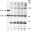
FIG. 1.
FIG. 1.
C3 binds to M. tuberculosis. M. tuberculosis bacilli (approximately 2.5 × 10⁷) were incubated in NHS for 5 to 60 min, cooled on ice, and then washed three times in ice-cold PBS containing protease inhibitors. Sample buffer was added to the washed bacterial pellets or purified C3 protein, the samples were heated to 100°C, resolved by SDS-7.5% PAGE, and transferred to nitrocellulose. Western blotting was performed with a polyclonal antibody to human C3 protein. Lane 1, 200 ng of purified C3 protein; lanes 2 to 6, M. tuberculosis bacilli incubated in NHS for 5, 10, 15, 30, and 60 min, respectively. The positions of C3α and C3β are shown on the left. The molecular mass markers are shown on the right. The results shown are representative of three independent experiments.
FIG. 2.
FIG. 2.
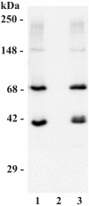
C3 binds to M. tuberculosis via the alternative pathway in 25% NHS. M. tuberculosis bacilli (2.5 × 10⁷) were incubated in NHS in the presence or absence of 10 mM EDTA or EGTA-Mg for 30 min at 37°C. The bacterial pellets were then washed and processed for Western blotting as described in the legend of Fig. 1. Lane 1, control condition in the presence of NHS without EDTA or EGTA; lane 2, EDTA; lane 3, EGTA-Mg. The molecular mass indicators are shown on the left. The results shown are representative of three independent experiments.
FIG. 3.
FIG. 3.
Hydroxylamine releases bound C3 from M. tuberculosis bacilli. M. tuberculosis bacilli (2.5 × 10⁷) were incubated in NHS at 37°C for 30 min and then washed three times in ice-cold PBS containing protease inhibitors. M. tuberculosis pellets were then treated with hydroxylamine as described in Materials and Methods. The bacterial pellets and hydroxylamine supernatants were then resolved by SDS-7.5% PAGE under reducing conditions and subjected to Western blotting with an anti-human C3 antibody. Lane 1, 200 ng of purified C3 protein; lane 2, NHS-incubated M. tuberculosis bacilli without hydroxylamine treatment; lane 3, M. tuberculosis pellet after hydroxylamine treatment; lane 4, hydroxylamine supernatant from the M. tuberculosis pellet in lane 3. The molecular mass indicators are shown on the left. The positions of C3b (arrowhead) and C3bi (arrow) are shown on the right. The results shown are representative of four independent experiments.
FIG. 4.
FIG. 4.
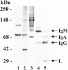
C3 binds multiple targets on M. tuberculosis. Purified C3 protein (200 ng; lane 1), washed NHS-incubated M. tuberculosis pellets (lanes 2 to 4), or 20 μl of NHS diluted 1/1,000 (lane 5), was resolved by SDS-7.5% PAGE under reducing conditions and then transferred to nitrocellulose. The lanes were divided, incubated in anti-C3 antibody (lanes 1 to 2), monoclonal anti-C3bi (lane 3), or anti-IgGAM antibody (lanes 4 to 5) and then the appropriate secondary antibody. The molecular mass markers are shown on the left. The positions of IgM, IgA, IgG, and light (L) chain are shown on the right. The results shown are representative of three independent experiments.
FIG. 5.
FIG. 5.
C3 binds to M. tuberculosis proteins. M. tuberculosis bacilli (2.5 × 10⁷) were incubated with NHS in the absence (lanes 1 and 3) or presence (lanes 2 and 4) of 10 mM EDTA, washed, resolved by SDS-PAGE under reducing conditions, and transferred to nitrocellulose. The lanes were divided and then incubated in anti-C3 antibody (lanes 1 and 2), or anti-M. tuberculosis antiserum (lanes 3 and 4). The results shown are representative of three independent experiments. The molecular mass markers are shown on the left.
FIG. 6.
FIG. 6.
C3 binding to M. tuberculosis is reduced in the absence of C1q. M. tuberculosis bacilli (2.5 × 10⁷) were incubated in 2.5% pooled human serum (PHS), 2.5% C1qD, 2.5% C1qD repleted with 200 μg of C1q protein per ml of serum (C1qD + C1q), FBD, or FBD repleted with 200 μg of FB per ml of serum (FBD + FB), for 30 min at 37°C. The bacterial pellets were washed, and C3 binding was detected by ELISA. Shown are the means ± standard errors of the means of triplicate determinations of four to seven independent experiments. *, P < 0.05.
FIG. 7.
FIG. 7.
C3 protein is detected in BAL fluid. Purified C3 protein (200, 100, 50, or 25 ng in lanes 1 to 4, respectively), 20 μl of BAL fluid (lane 5), or 20 μl of twofold serial dilutions of cBAL fluid (lanes 6 to 8) was resolved by SDS-PAGE under reducing conditions and subjected to Western blotting as described in the legend of Fig. 1. The molecular mass indicators are shown on the right. The positions of C3α and C3β are shown on the left. The results shown are representative of three independent experiments.
FIG. 8.
FIG. 8.
M. tuberculosis fixes C3 protein in BAL fluid. M. tuberculosis bacilli (2.5 × 10⁷) were incubated with 220 μl of cBAL fluid in the presence and absence of 10 mM EDTA or 10 mM EGTA-Mg for 30 min at 37°C, washed in ice-cold PBS containing protease inhibitors, and dispensed into triplicate wells of a 96-well microtiter plate. C3 binding was then determined by ELISA. The background (<0.05 A405) was equal to the results obtained in the presence of EDTA and was subtracted from the results shown. Shown are the means ± standard deviations of triplicate determinations from the three different donor cBAL fluid samples reported in Table 1 (where A is donor 1, B is donor 2, and C is donor 3). *, P < 0.05.
FIG. 9.
FIG. 9.
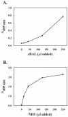
M. tuberculosis binds Ig in BAL fluid and NHS. M. tuberculosis bacilli (2.5 × 10⁷) were incubated with increasing amounts of cBAL fluid (A) or NHS (B) in a final volume of 500 μl as indicated for 30 min at 37°C, washed in ice-cold PBS containing protease inhibitors, and dispensed into triplicate wells of a 96-well microtiter plate and evaporated to dryness. The wells were then blocked, incubated in HRP anti-human IgGAM antibody, and developed with peroxidase substrate. The A405 was determined for each well, and the background (wells that did not contain M. tuberculosis; A405 ≤ 0.05) was subtracted out in each case. Shown are the means ± standard deviations of triplicate determinations from one experiment representative of three.

Similar articles

Cited by

References

    1. Actor, J. K., E. Breij, R. A. Wetsel, H. Hoffmann, R. L. Hunter, Jr., and C. Jagannath. 2001. A role for complement C5 in organism containment and granulomatous response during murine tuberculosis. Scand. J. Immunol. 53:464-474. - PubMed
    1. Armstrong, J. A., and P. D. Hart. 1971. Response of cultured macrophages to Mycobacterium tuberculosis, with observations on fusion of lysosomes with phagosomes. J. Exp. Med. 134:713-740. - PMC - PubMed
    1. Bardana, E. J. J., J. K. McClatchy, R. S. Farr, and P. Minden. 1973. Universal occurrence of antibodies to tubercle bacilli in sera from non-tuberculous and tuberculous individuals. Clin. Exp. Immunol. 13:65-77. - PMC - PubMed
    1. Berger, M. 1990. Third component of human complement: C3. Methods Enzymol. 184:619-628. - PubMed
    1. Bohlson, S. S., J. A. Strasser, J. J. Bower, and J. S. Schorey. 2001. Role of complement in Mycobacterium avium pathogenesis: in vivo and in vitro analyses of the host response to infection in the absence of complement component C3. Infect. Immun. 69:7729-7735. - PMC - PubMed

Publication types

MeSH terms