

Platelet aggregation induced by serotype polysaccharides from Streptococcus mutans
- PMID: 15102769
- PMCID: PMC387875
- DOI: 10.1128/IAI.72.5.2605-2617.2004
Platelet aggregation induced by serotype polysaccharides from Streptococcus mutans
Abstract
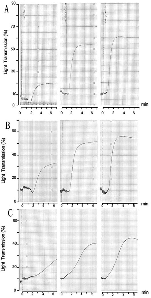
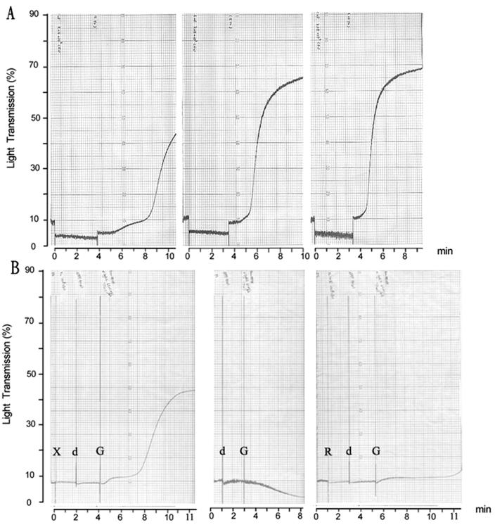
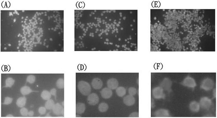
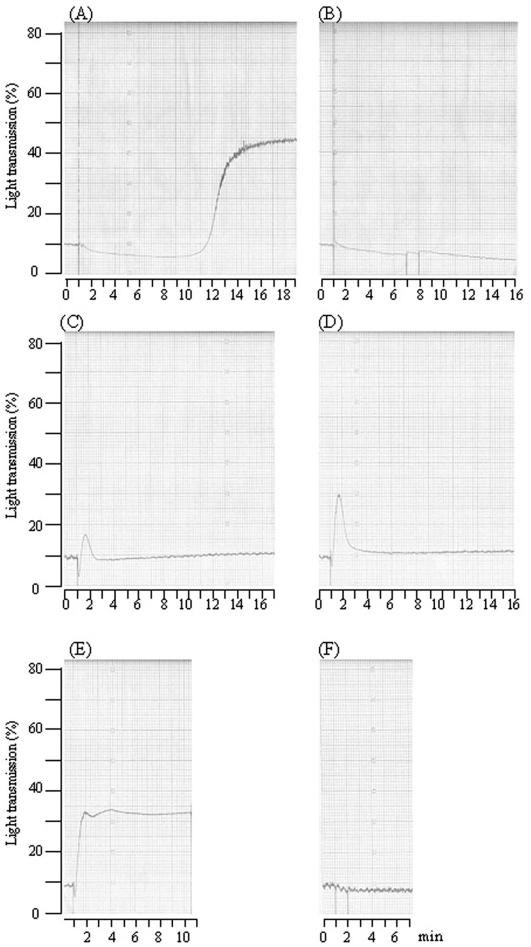
Platelet aggregation plays an important role in the pathogenesis of infective endocarditis induced by viridans streptococci or staphylococci. Aggregation induced in vitro involves direct binding of bacteria to platelets through multiple surface components. Using platelet aggregometry, we demonstrated in this study that two Streptococcus mutans laboratory strains, GS-5 and Xc, and two clinical isolates could aggregate platelets in an irreversible manner in rabbit platelet-rich plasma preparations. The aggregation was partially inhibited by prostaglandin I(2) (PGI(2)) in a dose-dependent manner. Whole bacteria and heated bacterial cell wall extracts were able to induce aggregation. Cell wall polysaccharides extracted from the wild-type Xc strain, containing serotype-specific polysaccharides which are composed of rhamnose-glucose polymers (RGPs), could induce platelet aggregation in the presence of plasma. Aggregation induced by the serotype-specific RGP-deficient mutant Xc24R was reduced by 50% compared to the wild-type strain Xc. In addition, cell wall polysaccharides extracted from Xc24R failed to induce platelet aggregation. The Xc strain, but not the Xc24R mutant, could induce platelet aggregation when preincubated with plasma. Both Xc and Xc24R failed to induce platelets to aggregate in plasma depleted of immunoglobulin G (IgG), but aggregation was restored by replenishment of anti-serotype c IgG. Analysis by flow cytometry showed that S. mutans RGPs could bind directly to rabbit and human platelets. Furthermore, cell wall polysaccharides extracted from the Xc, but not the Xc24R, strain could induce pseudopod formation of both rabbit and human platelets in the absence of plasma. Distinct from the aggregation of rabbit platelets, bacterium-triggered aggregation of human platelets required a prolonged lag phase and could be blocked completely by PGI(2). RGPs also trigger aggregation of human platelets in a donor-dependent manner, either as a transient and reversible or a complete and irreversible response. These results indicated that serotype-specific RGPs, a soluble product of S. mutans, could directly bind to and activate platelets from both rabbit and human. In the presence of plasma containing IgG specific to RGPs, RGPs could trigger aggregation of both human and rabbit platelets, but the degree of aggregation in human platelets depends on the donors.
Figures


Similar articles
-
Role of serotype-specific polysaccharide in the resistance of Streptococcus mutans to phagocytosis by human polymorphonuclear leukocytes.Infect Immun. 2000 Feb;68(2):644-50. doi: 10.1128/IAI.68.2.644-650.2000. Infect Immun. 2000. PMID: 10639428 Free PMC article.
-
Serotype-specific polysaccharide of Streptococcus mutans contributes to infectivity in endocarditis.Oral Microbiol Immunol. 2006 Dec;21(6):420-3. doi: 10.1111/j.1399-302X.2006.00317.x. Oral Microbiol Immunol. 2006. PMID: 17064403
-
Contribution of cell surface protein antigen c of Streptococcus mutans to platelet aggregation.Oral Microbiol Immunol. 2009 Oct;24(5):427-30. doi: 10.1111/j.1399-302X.2009.00521.x. Oral Microbiol Immunol. 2009. PMID: 19702959
-
Roles of oral bacteria in cardiovascular diseases--from molecular mechanisms to clinical cases: Cell-surface structures of novel serotype k Streptococcus mutans strains and their correlation to virulence.J Pharmacol Sci. 2010;113(2):120-5. doi: 10.1254/jphs.09r24fm. Epub 2010 May 24. J Pharmacol Sci. 2010. PMID: 20501965 Review.
-
Serotype classification of Streptococcus mutans and its detection outside the oral cavity.Future Microbiol. 2009 Sep;4(7):891-902. doi: 10.2217/fmb.09.64. Future Microbiol. 2009. PMID: 19722842 Review.
Cited by
-
The Bifidobacterium dentium Bd1 genome sequence reflects its genetic adaptation to the human oral cavity.PLoS Genet. 2009 Dec;5(12):e1000785. doi: 10.1371/journal.pgen.1000785. Epub 2009 Dec 24. PLoS Genet. 2009. PMID: 20041198 Free PMC article.
-
New insights into iron uptake in Streptococcus mutans: evidence for a role of siderophore-like molecules.Arch Microbiol. 2025 Mar 20;207(4):96. doi: 10.1007/s00203-025-04284-5. Arch Microbiol. 2025. PMID: 40111578
-
Sugar Allocation to Metabolic Pathways is Tightly Regulated and Affects the Virulence of Streptococcus mutans.Genes (Basel). 2016 Dec 28;8(1):11. doi: 10.3390/genes8010011. Genes (Basel). 2016. PMID: 28036052 Free PMC article. Review.
-
RgpF Is Required for Maintenance of Stress Tolerance and Virulence in Streptococcus mutans.J Bacteriol. 2017 Nov 14;199(24):e00497-17. doi: 10.1128/JB.00497-17. Print 2017 Dec 15. J Bacteriol. 2017. PMID: 28924033 Free PMC article.
-
Collagen-binding proteins of Streptococcus mutans and related streptococci.Mol Oral Microbiol. 2017 Apr;32(2):89-106. doi: 10.1111/omi.12158. Epub 2016 Apr 25. Mol Oral Microbiol. 2017. PMID: 26991416 Free PMC article. Review.
References
-
- Baddour, L. M. 1988. Twelve-year review of recurrent native valve infectious endocarditis: a disease of the modern antibiotic era. Rev. Infect. Dis. 10:1163-1170. - PubMed
-
- Bardsley, B., D. H. Williams, and T. P. Bablin. 1998. Cleavage of rhamnose from ristocetin A removes its ability to induce platelet aggregation. Blood Coagul. Fibrinolysis 9:241-244. - PubMed
-
- Braun, D. G. 1983. The use of streptococcal antigens to probe the mechanisms of immunity. Microbiol. Immunol. 27:823-836. - PubMed
Publication types
MeSH terms
Substances
LinkOut - more resources
Full Text Sources
Other Literature Sources
