. 2004 May;72(5):2723-30.
doi: 10.1128/IAI.72.5.2723-2730.2004.

Nitric oxide-mediated regulation of gamma interferon-induced bacteriostasis: inhibition and degradation of human indoleamine 2,3-dioxygenase

Affiliations

Nitric oxide-mediated regulation of gamma interferon-induced bacteriostasis: inhibition and degradation of human indoleamine 2,3-dioxygenase

Christian Hucke et al. Infect Immun. 2004 May.

Abstract

Tryptophan depletion resulting from indoleamine 2,3-dioxygenase (IDO) activity within the kynurenine pathway is one of the most prominent gamma interferon (IFN-gamma)-inducible antimicrobial effector mechanisms in human cells. On the other hand, nitric oxide (NO) produced by the inducible isoform of NO synthase (iNOS) serves a more immunoregulatory role in human cells and thereby interacts with tryptophan depletion in a number of ways. We investigated the effects of NO on IDO gene transcription, protein synthesis, and enzyme activity as well as on IDO-mediated bacteriostasis in the human epithelial cell line RT4. IFN-gamma-stimulated RT4 cells were able to inhibit the growth of Staphylococcus aureus in an IDO-mediated fashion, and this bacteriostatic effect was abolished by endogenously produced NO. These findings were supported by experiments which showed that IDO activity in extracts of IFN-gamma-stimulated cells is inhibited by the chemical NO donors diethylenetriamine diazeniumdiolate, S-nitroso-L-cysteine, and S-nitroso-N-acetyl-D,L-penicillamine. Furthermore, we found that both endogenous and exogenous NO strongly reduced the level of IDO protein content in RT4 cells. This effect was not due to a decrease in IDO gene transcription or mRNA stability. By using inhibitors of proteasomal proteolytic activity, we showed that NO production led to an accelerated degradation of IDO protein in the proteasome. This is the first report, to our knowledge, that demonstrates that the IDO is degraded by the proteasome and that NO has an effect on IDO protein stability.


Figures
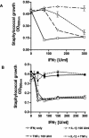
FIG. 1.
FIG. 1.
Inhibitory effect of IL-1β and TNF-α on IFN-γ-induced bacteriostasis in RT4 cells. We stimulated 3 × 10⁴ RT4 cells per well with IFN-γ alone or together with IL-1β and/or TNF-α. After 3 days of incubation, staphylococci were added (50 CFU/well). Bacterial growth was determined 16 h later by measuring the optical density at 600 nm (A). The same experimental procedure was performed in the presence of 100 μg of NG-monomethyl-l-arginine per ml. Data are given as mean optical density (OD) ± standard deviation for triplicate cultures (B). Addition of tryptophan (50 μg/ml) completely blocked IFN-γ induced bacteriostasis (B, solid symbols).
FIG. 2.
FIG. 2.
NO inhibits IDO activity in extracts of IFN-γ-stimulated cells. 86HG39 cells were stimulated with 300 U of IFN-γ per ml for 24 h. Per sample, 2 × 10⁶ cells were lysed, and the prepared cell extracts were assayed for IDO activity as described in Materials and Methods. The protein content of each sample was 0.312 μg. The NO donors DETA/NO, SNOC, and SNAP were added to the reaction buffer immediately before mixing with the cell extracts. Donor-NO represents the degassed (non-NO-producing) form of the donor. Results indicate the means ± standard error of the mean for three independent experiments done in duplicate. ***, P < 0.001, unpaired t test.
FIG. 3.
FIG. 3.
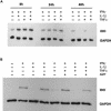
IDO protein content in RT4 cells decreases in a time- and NO-dependent manner. RT4 cells were stimulated for 8, 24, or 48 h with the indicated cytokines (each at 100 U/ml). Thereafter, cell extracts were prepared, 10 μg of total protein was separated by SDS-PAGE, and IDO protein was detected by Western blotting. As a quantity control, GAPDH was detected simultaneously (A). RT4 cells were stimulated for 48 h with the indicated cytokines (each at 100 U/ml) in the presence or absence of the selective iNOS inhibitor AMT (100 μg/ml). Thereafter, cells were lysed, and 10 μg of total protein was subjected to SDS-PAGE and Western blotting (B).
FIG. 4.
FIG. 4.
Quantification of IDO mRNA in RT4 cells under different stimulation conditions by real-time PCR. RT4 cells were stimulated with the indicated cytokines (each at 100 U/ml) for 8 h. Thereafter, RNA was extracted, and 1 μg of total RNA was used for reverse transcription. The resulting cDNA was amplified by real-time PCR as described in Materials and Methods. Standard solutions of a plasmid carrying the full-length IDO cDNA were used for quantification. Unstimulated cells and cells treated with cycloheximide (CHX, 10 μg/ml) or actinomycin D (ActD, 5 μg/ml) 1 h before addition of IFN-γ served as controls. The copy numbers of IDO mRNA were normalized to the number of GAPDH mRNA copies in the same probe. The values are the means ± standard error of the mean for the results of three independent experiments (A). There was no significant inhibition of IDO mRNA in cells treated with IFN-γ in the presence of IL-1 and/or TNF-α (P < 0.05, unpaired t test). iNOS activity in cell extracts of differently stimulated RT4 cells stimulated for 8 h with the indicated cytokines (each 100 U/ml) was determined. Cell extracts were prepared, and 100 μg of total protein per sample was assayed for iNOS activity with the NOSdetect assay kit as described in Materials and Methods. Cell extracts from unstimulated cells and samples containing the NOS inhibitor N-nitro-l-arginine (N-Nitro-Arg) served as internal controls as described in Materials and Methods. Data are given as means ± standard error of the mean for two independent experiments done in duplicate (B).
FIG. 5.
FIG. 5.
Effect of NO on IDO protein is not cycloheximide sensitive. A549 cells were stimulated with 50 U of IFN-γ per ml for 16 h. Thereafter, the cells were washed, and 10 μg of cycloheximide per ml was added in fresh culture medium. One hour later, the NO donor GSNO was added to a final concentration of 750 μM to some cells, and the cells were incubated for a further 4 to 24 h. Thereafter, cell extracts were prepared, and 10 μg of total protein was separated by SDS-PAGE. After blotting, IDO and GAPDH protein levels were determined.
FIG. 6.
FIG. 6.
Proteasome inhibitors abolish NO-dependent IDO degradation. A549 cells were stimulated with 50 U of IFN-γ per ml for 9 h. Thereafter, the cells were washed, and the proteasome inhibitors MG-132, proteasome inhibitor I (PSI), and clasto-lactacystin β-lactone (clasto) were added at the indicated final concentrations to fresh culture medium. The addition of dimethyl sulfoxide to a final concentration of 0.5% served as a solvent control (GSNO plus DMSO, lane 4), and unstimulated cells as a negative control (Med, lane 1). One hour later, the NO donor GSNO was added to a final concentration of 750 μM, and the cells were further incubated for 16 h. IDO and GAPDH protein levels were determined by Western blotting (A). The signal intensity of the IDO bands was measured by densitometry and normalized to the intensity of the corresponding GAPDH signal. The results shown are representative of one of three independent experiments (B).

Similar articles

Cited by

References

    1. Alberati-Giani, D., P. Malherbe, P. Ricciardi-Castagnoli, C. Köhler, S. Denis-Donini, and A. M. Cesura. 1997. Differential regulation of indoleamine-2,3-dioxygenase expression by nitric oxide and inflammatory mediators in IFN-γ-activated murine macrophages and microglial cells. J. Immunol. 159:419-426. - PubMed
    1. Bebok, Z., K. Varga, J. K. Hicks, C. J. Venglarik, T. Kovacs, L. Chen, K. M. Hardiman, J. F. Collawn, E. J. Sorscher, and S. Matalon. 2002. Reactive oxygen nitrogen species decrease cystic fibrosis transmembrane conductance regulator expression and cAMP-mediated Cl− secretion in airway epithelia. J. Biol. Chem. 277:43041-43049. - PubMed
    1. Bingisser, R. M., T. A. Tilbrook, P. G. Holt, and U. R. Kees. 1998. Macrophage-derived nitric oxide regulates T cell activation via reversible disruption of the Jak3/STAT5 signaling pathway. J. Immunol. 160:5729-5734. - PubMed
    1. Bogdan, C., M. Röllinghoff, and A. Diefenbach. 2000. Reactive oxygen and reactive nitrogen intermediates in innate and specific immunity. Curr. Opin. Immunol. 12:64-76. - PubMed
    1. Byrne, G. I., L. K. Lehmann, J. G. Kirschbaum, E. C. Borden, C. M. Lee, and R. R. Brown. 1986. Induction of tryptophan degradation in vitro and in vivo: a g-Interferon-stimulated activity. J. Interferon Res. 6:389-396. - PubMed

Publication types

MeSH terms