

Binding of hematin by a new class of glutathione transferase from the blood-feeding parasitic nematode Haemonchus contortus
- PMID: 15102788
- PMCID: PMC387910
- DOI: 10.1128/IAI.72.5.2780-2790.2004
Binding of hematin by a new class of glutathione transferase from the blood-feeding parasitic nematode Haemonchus contortus
Abstract
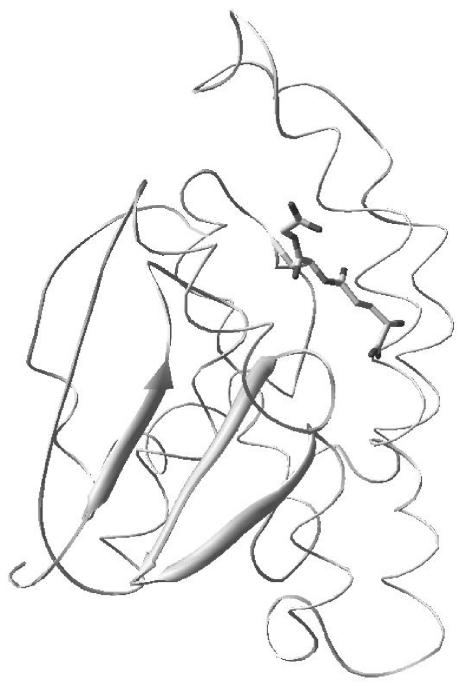
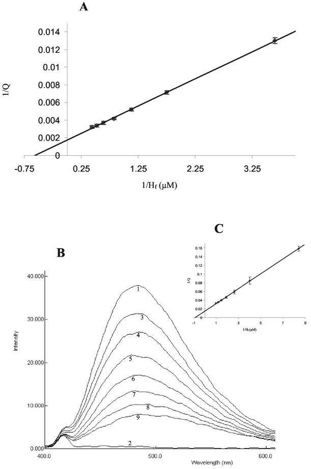
The phase II detoxification system glutathione transferase (GST) is associated with the establishment of parasitic nematode infections within the gastrointestinal environment of the mammalian host. We report the functional analysis of a GST from an important worldwide parasitic nematode of small ruminants, Haemonchus contortus. This GST shows limited activity with a range of classical GST substrates but effectively binds hematin. The high-affinity binding site for hematin was not present in the GST showing the most identity, CE07055 from the free-living nematode Caenorhabditis elegans. This finding suggests that the high-affinity binding of hematin may represent a parasite adaptation to blood or tissue feeding from the host.
Figures
Similar articles
-
Hc-hrg-2, a glutathione transferase gene, regulates heme homeostasis in the blood-feeding parasitic nematode Haemonchus contortus.Parasit Vectors. 2020 Jan 29;13(1):40. doi: 10.1186/s13071-020-3911-z. Parasit Vectors. 2020. PMID: 31996262 Free PMC article.
-
Molecular study of the transcription factor SKN-1 and its putative relationship with genes that encode GST and antioxidant enzymes in Haemonchus contortus.Vet Parasitol. 2024 Oct;331:110255. doi: 10.1016/j.vetpar.2024.110255. Epub 2024 Jul 14. Vet Parasitol. 2024. PMID: 39084102
-
Screening and analysis of Hc-ubq and Hc-gst related to desiccation survival of infective Haemonchus contortus larvae.Vet Parasitol. 2015 Jun 15;210(3-4):179-85. doi: 10.1016/j.vetpar.2015.03.020. Epub 2015 Mar 30. Vet Parasitol. 2015. PMID: 25913452
-
Atypical (RIO) protein kinases from Haemonchus contortus--promise as new targets for nematocidal drugs.Biotechnol Adv. 2011 May-Jun;29(3):338-50. doi: 10.1016/j.biotechadv.2011.01.006. Epub 2011 Jan 22. Biotechnol Adv. 2011. PMID: 21262337 Review.
-
Prospects for exploring molecular developmental processes in Haemonchus contortus.Int J Parasitol. 2006 Jul;36(8):859-68. doi: 10.1016/j.ijpara.2006.04.007. Epub 2006 May 17. Int J Parasitol. 2006. PMID: 16759659 Review.
Cited by
-
Proteomic profiling and protein identification by MALDI-TOF mass spectrometry in unsequenced parasitic nematodes.PLoS One. 2012;7(3):e33590. doi: 10.1371/journal.pone.0033590. Epub 2012 Mar 29. PLoS One. 2012. PMID: 22479418 Free PMC article.
-
Genome-wide analysis reveals novel genes essential for heme homeostasis in Caenorhabditis elegans.PLoS Genet. 2010 Jul 29;6(7):e1001044. doi: 10.1371/journal.pgen.1001044. PLoS Genet. 2010. PMID: 20686661 Free PMC article.
-
Altering the intracellular trafficking of Necator americanus GST-1 antigen yields novel hookworm mRNA vaccine candidates.PLoS Negl Trop Dis. 2025 Jan 10;19(1):e0012809. doi: 10.1371/journal.pntd.0012809. eCollection 2025 Jan. PLoS Negl Trop Dis. 2025. PMID: 39792959 Free PMC article.
-
Identification and characterisation of the haemozoin of Haemonchus contortus.Parasit Vectors. 2023 Mar 6;16(1):88. doi: 10.1186/s13071-023-05714-3. Parasit Vectors. 2023. PMID: 36879311 Free PMC article.
-
Safety and immunogenicity of the Na-GST-1 hookworm vaccine in Brazilian and American adults.PLoS Negl Trop Dis. 2017 May 2;11(5):e0005574. doi: 10.1371/journal.pntd.0005574. eCollection 2017 May. PLoS Negl Trop Dis. 2017. PMID: 28464026 Free PMC article. Clinical Trial.
References
-
- Barrett, J., N. Saghir, A. Timanova, K. Clarke, and P. M. Brophy. 1997. Characterisation and properties of an intracellular lipid-binding protein from the tapeworm Moniezia expansa. Eur. J. Biochem. 250:269-275. - PubMed
-
- Brophy, P. M., and J. Barrett. 1990. Strategies for detoxification of aldehydic products of lipid-peroxidation in helminths. Mol. Biochem. Parasitol. 42:205-211. - PubMed
-
- Brophy, P. M., A. Bensmith, A. Brown, J. M. Behnke, and D. I. Pritchard. 1995. Differential expression of glutathione-S-transferase (GST) by adult Heligmosomoides polygyrus during primary infection in fast and slow responding hosts. Int. J. Parasitol. 25:641-645. - PubMed
-
- Brophy, P. M., A. Bensmith, A. Brown, J. M. Behnke, and D. I. Pritchard. 1994. Glutathione S-transferases from the gastrointestinal nematode Heligmosomoides polygyrus and mammalian liver compared. Comp. Biochem. Physiol. Part B 109:585-592. - PubMed
-
- Brophy, P. M., A. Brown, and D. I. Pritchard. 1994. A PCR strategy for the isolation of glutathione S-transferases (GSTs) from nematodes. Int. J. Parasitol. 24:1059-1061. - PubMed
Publication types
MeSH terms
Substances
Associated data
- Actions
LinkOut - more resources
Full Text Sources
Research Materials
