. 2004 May;72(5):2791-802.
doi: 10.1128/IAI.72.5.2791-2802.2004.

Proteolytic processing of the Mycoplasma hyopneumoniae cilium adhesin

Affiliations

Proteolytic processing of the Mycoplasma hyopneumoniae cilium adhesin

Steven P Djordjevic et al. Infect Immun. 2004 May.

Abstract

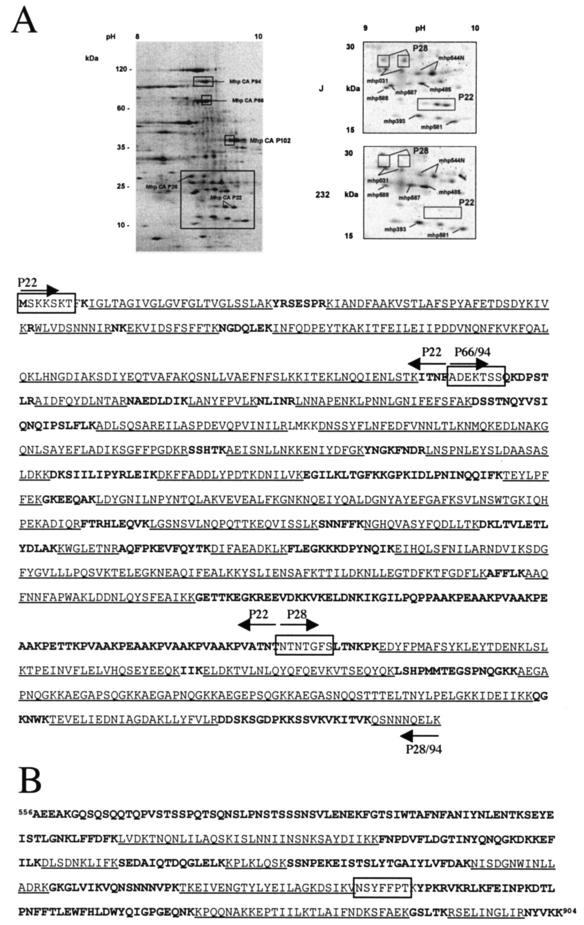
Mycoplasma hyopneumoniae is an economically significant swine pathogen that colonizes the respiratory ciliated epithelial cells. Cilium adherence is mediated by P97, a surface protein containing a repeating element (R1) that is responsible for binding. Here, we show that the cilium adhesin is proteolytically processed on the surface. Proteomic analysis of strain J proteins identified cleavage products of 22, 28, 66, and 94 kDa. N-terminal sequencing showed that the 66- and 94-kDa proteins possessed identical N termini and that the 66-kDa variant was generated by cleavage of the 28-kDa product from the C terminus. The 22-kDa product represented the N-terminal 195 amino acids of the cilium adhesin preprotein, confirming that the hydrophobic leader signal sequence is not cleaved during translocation across the membrane. Comparative studies of M. hyopneumoniae strain 232 showed that the major cleavage products of the cilium adhesin are similar, although P22 and P28 appear to be processed further in strain 232. Immunoblotting studies using antisera raised against peptide sequences within P22 and P66/P94 indicate that processing is complex, with cleavage occurring at different frequencies within multiple sites, and is strain specific. Immunogold electron microscopy showed that fragments containing the cilium-binding site remained associated with the cell surface whereas cleavage products not containing the R1 element were located elsewhere. Not all secreted proteins undergo multiple cleavage, however, as evidenced by the analysis of the P102 gene product. The ability of M. hyopneumoniae to selectively cleave its secreted proteins provides this pathogen with a remarkable capacity to alter its surface architecture.


Figures

FIG. 1.
FIG. 1.
Map of the cilium adhesin of M. hyopneumoniae and immunoblot analysis using MAb F1B6 and anti-peptide, P97 N-terminal, and R2 antisera. (A) The map shows antibody epitope locations, repeat regions, and selected cleavage sites identified by peptide mass fingerprint analysis, fragment masses, and N-terminal sequences. The major cleavage event at amino acid 195 (large arrow) and another event at amino acid 891 (small arrow) are shown. The locations of the R1 and R2 repeat regions are represented by gray-shaded boxes. The locations of the epitope for MAb F1B6, ΔNP97 peptide, and NP97, P97 N-terminal (P97 N-term), and P28 antisera are shown as bars above or below the map. The coiled-coil regions are represented by the black boxes. The cleavage products of strains J and 232 are shown as gray-shaded bars below the map. Their molecular masses and N-terminal sequences are shown to the right of the map. ND, not determined. *, the peptide TSSQKDPSTLR was identified by peptide mass fingerprinting P70 from strain 232, suggesting that this molecule has the same N-terminal sequence as P97. (B) Immunoblot patterns of M. hyopneumoniae strain J reacted with F1B6 (1:3,000), ΔNP97 (1:20), and NP97 (1:20). An SDS-12% polyacrylamide gel was used to resolve J strain cell lysates. Molecular mass markers (MBI Fermentas) are shown on the left. Equivalent amounts of M. hyopneumoniae cell lysate were loaded in lanes reacted with ΔNP97 (lane 2) and NP97 (lane 3) antibodies. The amount of protein loaded in lane 1 (reacted with MAb F1B6) was approximately one-third of that loaded in lanes 2 and 3. (C) Immunoblot patterns (12% polyacrylamide gel) of synchronized M. hyopneumoniae strain J cultures harvested at different times postinoculation. The blot was reacted with MAb F1B6 (1:1,000). Equivalent amounts of protein were loaded in each lane. Lane 1, 8 h of growth (late lag phase); lane 2, 16 h of growth (early exponential phase); lane 3, 20 h of growth (mid-log phase); lane 4, 24 h of growth (mid-log phase); lane 5, 28 h of growth (mid-log phase); lane 6, 40 h of growth (late log-early stationary phase); lane 7, 48 h of growth (stationary phase); lane 8, 52 h of growth (stationary phase); lane 9, 56 h of growth (stationary phase). (D) Immunoblot patterns (12% polyacrylamide gel) of synchronized M. hyopneumoniae strain J (left panel) and 232 (right panel) cultures harvested at different times postinoculation. The blot was reacted with P97 N-terminal serum (1:100). Equivalent amounts of protein were loaded in each lane. Lane 1, 20 h of growth (mid-log phase); lane 2, 28 h of growth (mid log phase); lane 3, 40 h of growth (late log-early stationary phase); lane 4, 56 h of growth (stationary phase). (E) Immunoblot patterns (10% polyacrylamide gel) of synchronized M. hyopneumoniae strain J (left panel) and 232 (right panel) cultures harvested at different times postinoculation. The blot was reacted with R2 serum (1:100). Equivalent amounts of protein were loaded in each lane. (Left panel) Lane 1, 8 h of growth (late log phase); lane 2, 16 h of growth (early exponential phase); lane 3, 20 h of growth (mid-log phase); lane 4, 24 h of growth (mid-log phase); lane 5, 28 h of growth (mid-log phase); lane 6, 40 h of growth (late log-early stationary phase); lane 7, 48 h of growth (stationary phase). (Right panel) Lane 1, 8 h of growth (late log phase); lane 2, 16 h of growth (early exponential phase); lane 3, 24 h of growth (mid-log phase); lane 4, 28 h of growth (mid-log phase); lane 5, 32 h (mid-log phase); lane 6, 40 h of growth (late log-early stationary phase); lane 7, 48 h of growth (stationary phase). (F) Comparison (using NP97 and ΔNP97 antisera) of immunoblot patterns of strains 232 and J. Molecular weight markers (Bio-Rad Precision Proteins Standards) (broad range) are shown on the right of the panel. An 8 to 16% Bio-Rad Criterion gradient gel was used to separate the proteins.
FIG. 2.
FIG. 2.
Peptide mass fingerprint analysis of the cilium adhesin. (A) (Upper panel) 2-D electrophoresis was used to resolve M. hyopneumoniae proteins, which were subsequently analyzed by peptide mass fingerprinting. (Upper left panel) Analysis of total J strain proteins. A region of the gel representing pH 8 to 10 is shown. The upper boxes indicate the proportion of the spots containing either P94J, P66J, or the 42-kDa fragment of P102 as indicated. The lower larger boxed area is shown in expanded form on the right for a comparison of strains J and 232 in the region of P28. (Lower panel) MALDI-TOF (MS) was used to analyze tryptic digests of indicated spots. The resulting fingerprints were matched to a database containing theoretical tryptic digests of M. hyopneumoniae ORFs derived from genome sequencing analysis (Minion, unpublished). Each cleavage product (P22, P28, P66, and P94) is shown by the arrows above and below the sequence. Underlined sequences were matched by MALDI-TOF (MS). Unmatched sequences are shown in bold characters. Boxed sequences represent N-terminal protein sequences of isolated spots obtained by Edman degradation. (B) Peptide mass mapping of a 42-kDa C-terminal fragment of P102. Underlined sequences were matched by MALDI-TOF (MS). Unmatched sequences are boldface. ESI MS-MS analysis was used to identify the sequence NSYFFPT (underlined). The N-terminal sequence AEEAKG indicates the beginning of the 42-kDa P102 fragment.
FIG. 3.
FIG. 3.
Growth medium does not contain recombinant P97232 cleavage activity. Recombinant P97232 was incubated in fresh and spent media overnight, and the products were resolved by immunoblotting using MAb F1B6. Lanes: 1, molecular weight standards; 2, fresh medium; 3, spent medium; 4, fresh medium plus recombinant P97232; 5, spent medium plus recombinant P97232. The arrow indicates the position of purified recombinant P97232.
FIG. 4.
FIG. 4.
Trypsin digestion of M. hyopneumoniae strain J cells and immunoblotting with MAb F1B6 (1:5,000). Each lane contains approximately 10 μg of J strain protein. Approximately 50 mg of mycoplasma whole-cell protein was treated with the indicated concentration (in micrograms per milliliter) of trypsin for 15 min at 37°C. Molecular mass markers (Bio-Rad) are shown on the left. Lanes: 1, 0 μg/ml; 2, 0.3 μg/ml; 3, 0.5 μg/ml; 4, 1 μg/ml; 5, 3 μg/ml; 6, 10 μg/ml; 7, 50 μg/ml; 8, 300 μg/ml.
FIG. 5.
FIG. 5.
Immunolocalization of the cilium adhesin and cleavage products on the surface of M. hyopneumoniae strain 232. Thin sections were successively labeled with normal mouse (A), mouse MAb F1B6 (B and C), mouse anti-peptide ΔNP97 (D to F), or mouse anti-peptide NP97 (G and H) sera and 10-nm-diameter colloidal gold-conjugated goat anti-mouse Ig and negatively stained with 1% phosphotungstic acid. Arrows indicate gold particles. Bars, 1 μm.
FIG. 6.
FIG. 6.
Analysis of the surface protein P102 by immunoblotting and immunoelectron microscopy. (A) Analysis of recombinant P102 with a Coomassie blue-stained SDS-PAGE gel. Lane 1, uninduced E. coli culture; lane 2, induced E. coli culture; lane 3, purified polyHis-P102 fusion. Molecular weight markers (in thousands) are shown on the left. (B) Immunoblot of M. hyopneumoniae whole-cell lysate with anti-P102 antiserum. Molecular weight markers are shown on the left. (C) Immunoelectron microscopy of M. hyopneumoniae cells with anti-P102 antiserum. Bar, 1 μm.
FIG. 7.
FIG. 7.
Adhesin cleavage products identified by peptide mass mapping and N-terminal sequence analysis and those predicted from immunoblotting studies. Large arrows identify confirmed cleavage sites; smaller arrows indicate predicted but unconfirmed cleavage events. The locations of the epitopes for MAb F1B6, peptide ΔNP97 and NP97 antisera, and P97 N-terminal (P97 N-term) and P28 antisera are shown as black bars.

References

    1. Calcutt, M. J., M. F. Kim, A. B. Karpas, P. F. Muhlradt, and K. S. Wise. 1999. Differential posttranslational processing confers intraspecies variation of a major surface lipoprotein and a macrophage-activating lipopeptide of Mycoplasma fermentans. Infect. Immun. 67:760-771. - PMC - PubMed
    1. Ciprian, A., C. Pijoan, T. Cruz, J. Camacho, J. Tortora, G. Colmenares, R. R. Lopez, and M. de la Garza. 1988. Mycoplasma hyopneumoniae increases the susceptibility of pigs to experimental Pasteurella multocida pneumonia. Can. J. Vet. Res. 52:434-438. - PMC - PubMed
    1. Cordwell, S. J., D. J. Basseal, B. Bjellqvist, D. C. Shaw, and I. Humphery-Smith. 1997. Characterisation of basic proteins from Spiroplasma melliferum using novel immobilised pH gradients. Electrophoresis 18:1393-1398. - PubMed
    1. Coutte, L., E. Willery, R. Antoine, H. Drobecq, C. Locht, and F. Jacob-Dubuisson. 2003. Surface anchoring of bacterial subtilisin important for maturation function. Mol. Microbiol. 49:529-539. - PubMed
    1. Davis, K. L., and K. S. Wise. 2002. Site-specific proteolysis of the MALP-404 lipoprotein determines the release of a soluble selective lipoprotein-associated motif-containing fragment and alteration of the surface phenotype of Mycoplasma fermentans. Infect. Immun. 70:1129-1135. - PMC - PubMed

Publication types

MeSH terms