

Clostridium difficile toxin A carboxyl-terminus peptide lacking ADP-ribosyltransferase activity acts as a mucosal adjuvant
- PMID: 15102793
- PMCID: PMC387895
- DOI: 10.1128/IAI.72.5.2827-2836.2004
Clostridium difficile toxin A carboxyl-terminus peptide lacking ADP-ribosyltransferase activity acts as a mucosal adjuvant
Abstract
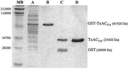
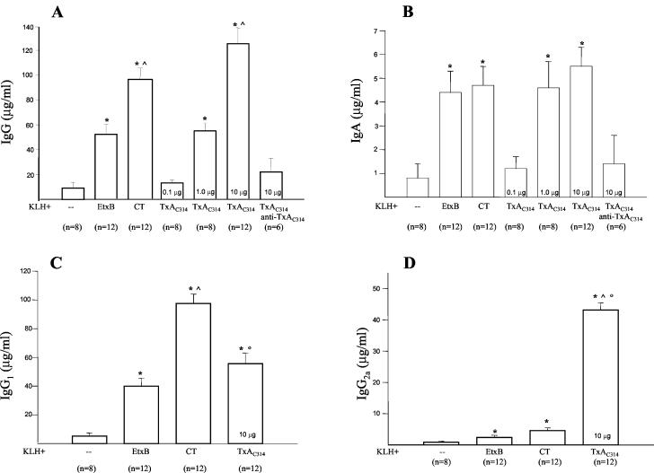
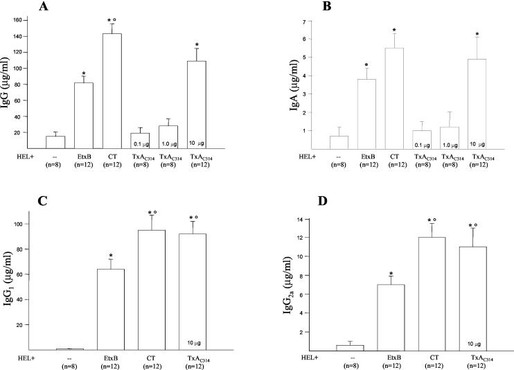
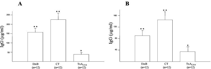
The receptor binding domains of the most potent mucosal adjuvants, bacterial toxins and plant lectins, are organized in repeat units to recognize specific sugar residues. The lectin-like structure of the C-terminal region of Clostridium difficile toxin A prompted us to investigate the mucosal adjuvant properties of a nontoxigenic peptide corresponding to amino acids 2394 to 2706 (TxA(C314)). We compared TxA(C314) adjuvant activity to those of cholera toxin (CT) and Escherichia coli heat-labile enterotoxin subunit B (EtxB) coadministered orally or nasotracheally with poor peptide antigens (keyhole limpet hemocyanin [KLH] and hen egg lysozyme [HEL]). Levels of anti-KLH-specific serum immunoglobulin G (IgG) and IgA as well as that of mucosal IgA were significantly higher in animals immunized orally with TxA(C314) plus KLH than with KLH alone, CT plus KLH, or EtxB plus KLH. Following intranasal immunization with TxA(C314) plus HEL, levels of serum- and mucosa-specific antibodies were comparable to those induced by coadministering HEL with CT or EtxB. The TxA(C314) adjuvant effect following oral, but not intranasal, immunization was dose dependent. The analysis of the subclasses of anti-KLH-specific IgG isotypes and the cytokines released from splenocytes of immunized mice challenged in vitro with KLH indicates the induction of a mixed Th1/Th2-type immune response, with prevalence of the Th1 branch. We conclude that TxA(C314) enhances immune responses against mucosa-coadministered foreign antigens and represents a promising mucosal adjuvant, especially because its ability to stimulate mixed Th1/Th2 responses with a strong a Th1 component is extremely worthwhile against intracellular pathogens.
Figures
Similar articles
-
The adjuvant effect of Vibrio cholerae and Escherichia coli heat-labile enterotoxins is linked to their ADP-ribosyltransferase activity.Eur J Immunol. 1992 Sep;22(9):2277-81. doi: 10.1002/eji.1830220915. Eur J Immunol. 1992. PMID: 1381311
-
Genetically detoxified mutants of heat-labile toxin from Escherichia coli are able to act as oral adjuvants.Infect Immun. 1999 Sep;67(9):4400-6. doi: 10.1128/IAI.67.9.4400-4406.1999. Infect Immun. 1999. PMID: 10456880 Free PMC article.
-
Comparative analysis of the mucosal adjuvanticity of the type II heat-labile enterotoxins LT-IIa and LT-IIb.Infect Immun. 2000 Jan;68(1):281-7. doi: 10.1128/IAI.68.1.281-287.2000. Infect Immun. 2000. PMID: 10603399 Free PMC article.
-
The role of ADP-ribosylation and G(M1)-binding activity in the mucosal immunogenicity and adjuvanticity of the Escherichia coli heat-labile enterotoxin and Vibrio cholerae cholera toxin.Immunol Cell Biol. 1998 Jun;76(3):270-9. doi: 10.1046/j.1440-1711.1998.00745.x. Immunol Cell Biol. 1998. PMID: 9682971 Review.
-
Immunomodulation using bacterial enterotoxins.Scand J Immunol. 2001 Mar;53(3):218-26. doi: 10.1046/j.1365-3083.2001.00884.x. Scand J Immunol. 2001. PMID: 11251877 Review.
Cited by
-
Nanobodies against C. difficile TcdA and TcdB reveal unexpected neutralizing epitopes and provide a toolkit for toxin quantitation in vivo.PLoS Pathog. 2023 Oct 23;19(10):e1011496. doi: 10.1371/journal.ppat.1011496. eCollection 2023 Oct. PLoS Pathog. 2023. PMID: 37871122 Free PMC article.
-
Transcutaneous immunization with Clostridium difficile toxoid A induces systemic and mucosal immune responses and toxin A-neutralizing antibodies in mice.Infect Immun. 2007 Jun;75(6):2826-32. doi: 10.1128/IAI.00127-07. Epub 2007 Mar 19. Infect Immun. 2007. PMID: 17371854 Free PMC article.
-
C-terminal repeats of Clostridium difficile toxin A induce production of chemokine and adhesion molecules in endothelial cells and promote migration of leukocytes.Infect Immun. 2008 Mar;76(3):1170-8. doi: 10.1128/IAI.01340-07. Epub 2007 Dec 26. Infect Immun. 2008. PMID: 18160482 Free PMC article.
-
A novel multivalent, single-domain antibody targeting TcdA and TcdB prevents fulminant Clostridium difficile infection in mice.J Infect Dis. 2014 Sep 15;210(6):964-72. doi: 10.1093/infdis/jiu196. Epub 2014 Mar 27. J Infect Dis. 2014. PMID: 24683195 Free PMC article.
-
Adenovirus-based vaccination against Clostridium difficile toxin A allows for rapid humoral immunity and complete protection from toxin A lethal challenge in mice.Vaccine. 2012 Feb 14;30(8):1492-501. doi: 10.1016/j.vaccine.2011.12.064. Epub 2011 Dec 23. Vaccine. 2012. PMID: 22200503 Free PMC article.
References
-
- Castagliuolo, I., A. Keates, C. Wang, A. Pasha, L. Valenick, C. P. Kelly, S. Nikulasson, J. T. LaMont, and C. Pothoulakis. 1998. Clostridium difficile toxin A stimulates macrophage inflammatory protein-2 production in rat intestinal epithelial cells. J. Immunol. 160:6039-6045. - PubMed
-
- Castagliuolo, I., and J. T. LaMont. 1999. Pathophysiology, diagnosis and treatment of Clostridium difficile infection. Keio J. Med. 48:169-174. - PubMed
-
- Clark, M. A., M. A. Jepson, N. L. Simmons, T. A. Booth, and B. H. Hirst. 1993. Differential expression of lectin-binding sites defines mouse intestinal M-cells. J. Histochem. Cyochem. 41:1679-1687. - PubMed
MeSH terms
Substances
LinkOut - more resources
Full Text Sources
Other Literature Sources
Miscellaneous
