. 2004 May;72(5):2864-71.
doi: 10.1128/IAI.72.5.2864-2871.2004.

Borrelia burgdorferi-induced expression of matrix metalloproteinases from human chondrocytes requires mitogen-activated protein kinase and Janus kinase/signal transducer and activator of transcription signaling pathways

Affiliations

Borrelia burgdorferi-induced expression of matrix metalloproteinases from human chondrocytes requires mitogen-activated protein kinase and Janus kinase/signal transducer and activator of transcription signaling pathways

Aruna K Behera et al. Infect Immun. 2004 May.

Abstract

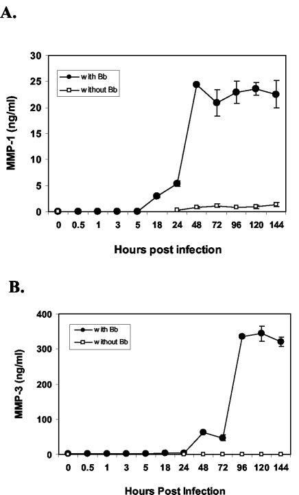
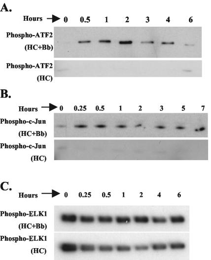
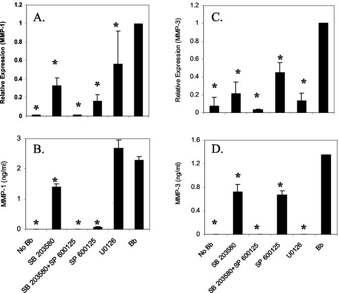
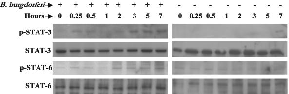
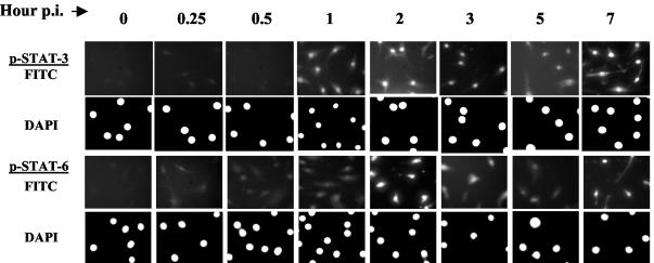
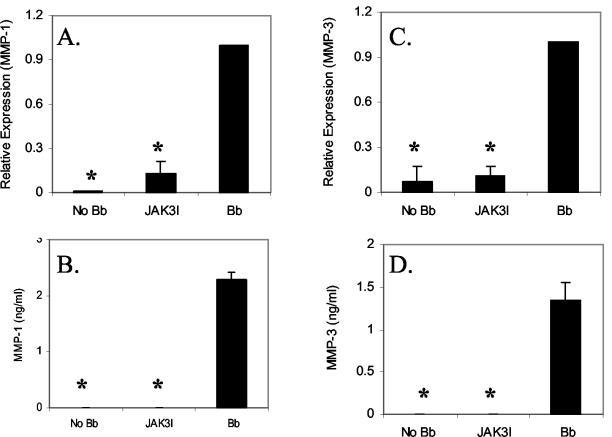
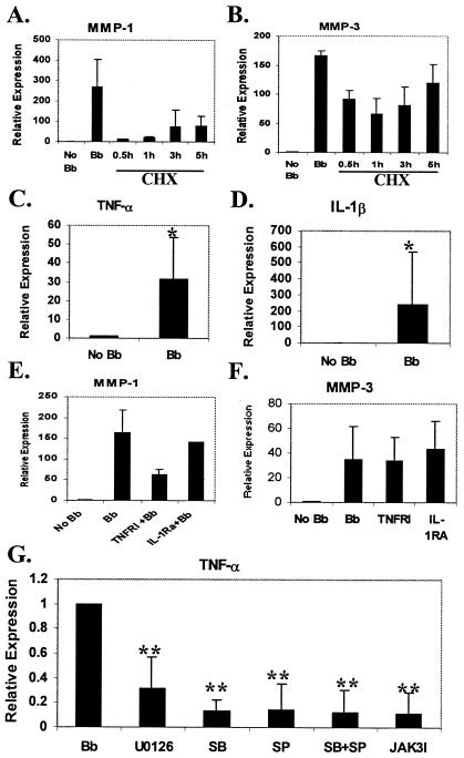
Elevations in matrix metalloproteinase 1 (MMP-1) and MMP-3 have been found in patients with Lyme arthritis and in in vitro models of Lyme arthritis using cartilage explants and chondrocytes. The pathways by which B. burgdorferi, the causative agent of Lyme disease, induces the production of MMP-1 and MMP-3 have not been elucidated. We examined the role of the extracellular signal-regulated kinase 1/2 (ERK1/2), c-Jun N-terminal kinase (JNK), p38 mitogen-activated protein kinase (MAPK) and the Janus kinase/signal transducer and activator of transcription (JAK/STAT) pathways in MMP induction by B. burgdorferi. Infection with B. burgdorferi results in rapid phosphorylation of p38 and JNK within 15 to 30 min. Inhibition of JNK and p38 MAPK significantly reduced B. burgdorferi-induced MMP-1 and MMP-3 expression. Inhibition of ERK1/2 completely inhibited the expression of MMP-3 in human chondrocytes following B. burgdorferi infection but had little effect on the expression of MMP-1. B. burgdorferi infection also induced phosphorylation and nuclear translocation of STAT-3 and STAT-6 in primary human chondrocytes. Expression of MMP-1 and MMP-3 was significantly inhibited by inhibition of JAK3 activity. Induction of MMP-1 and -3 following MAPK and JAK/STAT activation was cycloheximide sensitive, suggesting synthesis of intermediary proteins is required. Inhibition of tumor necrosis factor alpha (TNF-alpha) significantly reduced MMP-1 but not MMP-3 expression from B. burgdorferi-infected cells; inhibition of interleukin-1beta (IL-1beta) had no effect. Treatment of B. burgdorferi-infected cells with JAK and MAPK inhibitors significantly inhibited TNF-alpha induction, consistent with at least a partial role for TNF-alpha in B. burgdorferi-induced MMP-1 expression in chondrocytes.


Figures

FIG. 1.
FIG. 1.
Expression of MMP-1 and MMP-3 in B. burgdorferi (Bb)-infected human primary chondrocytes. Primary HC cultures were either uninfected or infected with B. burgdorferi (107 organisms) for various lengths of time. Expression of MMP-1 (A) and MMP-3 (B) was measured by ELISA. The experiment was repeated twice in duplicate and a representative experiment is shown. Error bars represent standard deviations.
FIG. 2.
FIG. 2.
Activation of ERK1/2, p38 MAPK, and JNK in human primary chondrocytes following B. burgdorferi infection. Primary HC cultures were infected with B. burgdorferi (Bb) for various lengths of time. Uninfected HCs harvested at the same time points were used as controls (lower panels). All cellular lysates were normalized to a total of 200 μg of protein prior to performing immunoprecipitation. (A) Activity of p38 MAPK was measured by immunoprecipitation of phospho-p38 MAPK, determining its activity by its ability to phosphorylate its substrate ATF-2. (B) Activity of JNK MAPK was measured by immunoprecipitating phospho-JNK, determining its activity by its ability to phosphorylate its substrate c-Jun. (C) The activity of ERK1/2 was measured by immunoprecipitation of phospho-ERK1/2, determining its kinase activity by phosphorylation of its substrate ELK-1. The experiments were repeated three to five times and representative experiments are shown.
FIG. 3.
FIG. 3.
Role of p38 MAPK, ERK1/2, and JNK on B. burgdorferi-induced MMP-1 and MMP-3 expression in primary HCs. Primary HC cultures were treated either with p38 MAPK inhibitor (SB203580); JNK inhibitor (SP600125); or ERK1/2 inhibitor (U0126) for 2 h and subsequently infected with B. burgdorferi. Cells were harvested at 18 hpi and MMP-1 and -3 expression was examined by real-time RT-PCR (A and C) and by ELISA (B and D). The real-time RT-PCR experiments were repeated three to five times and the ELISAs were repeated twice in duplicate. Error bars represent standard deviations. *, P < 0.05.
FIG. 4.
FIG. 4.
Activation of STAT molecules in primary HCs following B. burgdorferi infection. Primary HC cultures were either uninfected or infected with B. burgdorferi for various amounts of time and whole cell protein extracts were analyzed by immunoblotting for the expression of phospho-STAT-3 and phospho-STAT-6 using specific antibodies. Immunoblots for nonphosphorylated STAT-3 and STAT-6 are shown as internal controls. Experiments were performed three times and representative experiments are shown.
FIG. 5.
FIG. 5.
Nuclear translocation of phospho-STAT molecules in HC following B. burgdorferi infection. Primary HC were infected with B. burgdorferi for various periods of time. Nuclear translocation of STAT-3 and STAT-6 was examined by immunofluorescence using phospho-specific antibodies to individual STAT proteins and subsequently probed with FITC-labeled secondary antibody. The nucleus was counter stained with DAPI and photographed under fluorescence microscopy. The upper and lower panels show FITC and DAPI stain, respectively, for each of the STAT proteins. Experiments were performed two to three times and representative experiments are shown.
FIG. 6.
FIG. 6.
Role of JAK/STAT pathway on B. burgdorferi-induced MMP-1 and MMP-3 expression in primary HCs. HCs were treated with JAK3 inhibitor (JAK3I) for 2 h followed by infection with B. burgdorferi. Cells were harvested at 18 hpi and the expression of MMP-1 and -3 was examined by real-time PCR (A and C) and ELISA (B and D). The real-time RT-PCR experiments were repeated three times and the ELISAs were repeated twice. Bar represents standard deviation. *, P < 0.05.
FIG. 7.
FIG. 7.
Mechanism of activation of MMP-1 and MMP-3 following MAPK and STAT activation. HC were treated with CHX (10 μg/ml) at different time point as indicated following B. burgdorferi infection. Cells were harvested at 24 hpi and real-time RT-PCR was performed for MMP-1 (A) and MMP-3 (B). (C and D) Real-time RT-PCR analysis of TNF-α (C) and IL-1β (D) expression in HC following 24 h of B. burgdorferi infection. (E and F) Effect of inhibition of TNF-α and IL-1β expression by TNFRI and IL-1RA on MMP-1 and MMP-3 expression. HC were treated with TNFRI (2 μg/ml) and of IL-1RA (1 μg/ml) for 2 h and subsequently infected with B. burgdorferi. Cells were harvested at 24 hpi and real-time RT-PCR was done examine the expression of MMP-1 (E) and MMP-3 (F). (G) Effect of inhibitors of MAPKs and JAK/STAT pathways on TNF-α expression. HC were treated either with p38 MAPK inhibitor (SB) SB203580; JNK inhibitor (SP) SP600125; ERK1/2 inhibitors (U0126), or JAK3 inhibitor (JAK3I) for 2 h and subsequently infected with B. burgdorferi. Cells were harvested at 18 hpi, and the expression of MMP-1 and -3 was examined by real-time PCR. All the experiments were repeated two to four times in duplicate. Bar represents standard errors of the means. Symbols: *, P < 0.05; **, P < 0.01.

Similar articles

Cited by

References

    1. Barbour, A. 1984. Isolation and cultivation of Lyme disease spirochetes. Yale J. Biol. Med. 57:521-525. - PMC - PubMed
    1. Beck, G., J. L. Benach, and G. S. Habicht. 1989. Isolation of interleukin 1 from joint fluids of patients with Lyme disease. J. Rheumatol. 16:800-806. - PubMed
    1. Borzì, R. M., I. Mazzetti, L. Cattini, M. Uguccioni, M. Baggiolini, and A. Facchini. 2000. Human chondrocytes express functional chemokine receptors and release matrix-degrading enzymes in response to C-X-C and C-C chemokines. Arthritis Rheum. 43:1734-1741. - PubMed
    1. Chang, L., and M. Karin. 2001. Mammalian MAP kinase signalling cascades. Nature 410:37-40. - PubMed
    1. Coleman, J. L., T. J. Sellati, J. E. Testa, R. R. Kew, M. B. Furie, and J. L. Benach. 1995. Borrelia burgdorferi binds plasminogen, resulting in enhanced penetration of endothelial monolayers. Infect. Immun. 63:2478-2484. - PMC - PubMed

Publication types

MeSH terms