. 2004 May;72(5):2872-8.
doi: 10.1128/IAI.72.5.2872-2878.2004.

Mycobacteria inhibit nitric oxide synthase recruitment to phagosomes during macrophage infection

Affiliations

Mycobacteria inhibit nitric oxide synthase recruitment to phagosomes during macrophage infection

Barbara H Miller et al. Infect Immun. 2004 May.

Abstract

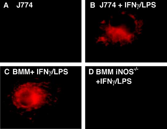
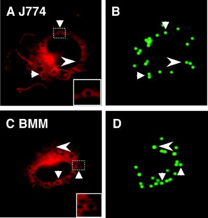
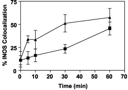
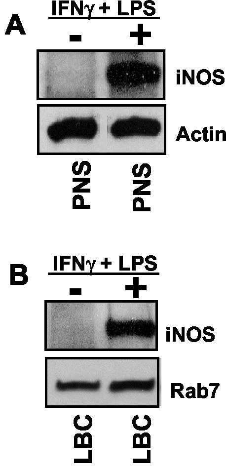
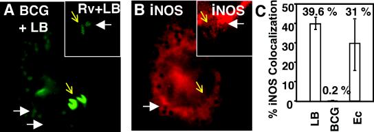
Inducible nitric oxide synthase (iNOS) is a cytoplasmic protein responsible for the generation of nitric oxide (NO. ) in macrophages. In this work, we hypothesized that the intracellular localization of iNOS is significant for effective delivery of NO. to phagosomes containing ingested microorganisms. Using immunofluorescence microscopy and Western blot analysis, iNOS was shown to localize in the vicinity of phagosomes containing latex beads in stimulated macrophages. iNOS also localized to phagosomes containing Escherichia coli. The colocalization of iNOS with ingested latex beads was an actin-dependent process, since treatment with the actin microfilament disrupter cytochalasin D prevented iNOS recruitment to latex bead phagosomes. In contrast to E. coli and inert particle phagosomes, mycobacterial phagosomes did not colocalize with iNOS. This study demonstrates that (i). iNOS can be recruited to phagosomes; (ii). this recruitment is dependent on a functional actin cytoskeleton; (iii). certain microorganisms have the ability to prevent or reduce colocalization with iNOS; and (iv). spatial exclusion of iNOS may play a role in Mycobacterium tuberculosis pathogenesis.


Figures

FIG. 1.
FIG. 1.
iNOS detection in macrophages by immunofluorescence microscopy. Images show the level of fluorescence observed with resting J774 cells (A), IFN-γ- and LPS-stimulated J774 macrophages (B), IFN-γ- and LPS-stimulated BMM from C57BL/6 mice (C), and IFN-γ- and LPS-stimulated BMM from C57BL/6 iNOS−/− mice (D). Magnification, ×100.
FIG. 2.
FIG. 2.
Recruitment of iNOS to model, LB phagosomes: J774 macrophages (A and B) and BMM from C57BL/6 mice (C and D) stimulated with IFN-γ and LPS. After phagocytosis of LB, cells were stained with anti-iNOS antibodies followed by a secondary antibody conjugated to Alexa 568. Arrows indicate examples of iNOS colocalization with LB phagosomes. Arrowheads indicate LB phagosomes that did not colocalize with iNOS. Insets (A and D) are enlarged images corresponding to the dashed boxes, as indicated.
FIG. 3.
FIG. 3.
Quantitative analysis of iNOS colocalization with phagosomes. Colocalization of LB phagosomes and iNOS over time in IFN-γ- and LPS-stimulated J774 macrophages and in IFN-γ- and LPS-stimulated BMM from C57BL/6 mice. Each symbol represents the mean percent iNOS colocalization for at least four microscopic fields containing a total of at least 50 LB phagosomes. Squares, J774; triangles, BMM.
FIG. 4.
FIG. 4.
Biochemical analysis of iNOS recruitment to purified phagosomes. (A) Western blot of PNS from unstimulated (−) or IFN-γ- and LPS-stimulated (+) J774 macrophages used to purify LBC. (B) Western blot of LBC purified from unstimulated (−) or IFN-γ- and LPS-stimulated (+) J774 macrophages. Blots were probed with antibodies to iNOS and actin (A) or iNOS and Rab7 (B). Actin and Rab7 served as loading controls for PNS and LBC, respectively.
FIG. 5.
FIG. 5.
Exclusion of iNOS from mycobacterial phagosomes in IFN-γ- and LPS-stimulated macrophages infected with LB, M. bovis BCG, or M. tuberculosis H37Rv. (A) Fluorescence (green) indicates LB, M. bovis BCG, or M. tuberculosis H37Rv; (B) fluorescence (red) indicates iNOS. The large panels show BCG-LB coinfection. The insets show macrophages coinfected with M. tuberculosis H37Rv (Rv) and LB (images are representative of three independent experiments). White arrows, examples of iNOS recruitment to LB phagosomes; yellow arrows, M. bovis BCG or M. tuberculosis H37Rv (expressing GFP). Note that iNOS did not colocalize with M. bovis BCG or M. tuberculosis H37Rv. (C) LB, M. bovis BCG, and E. coli DH5α (Ec) in IFN-γ- and LPS-stimulated J774 macrophages were scored for percent iNOS colocalization at 1 h postinfection.
FIG. 6.
FIG. 6.
A functional actin cytoskeleton is necessary for iNOS recruitment to phagosomes. Quantitation of iNOS colocalization with LB phagosomes in IFN-γ- and LPS-stimulated J774 macrophages under conditions of no treatment, cytochalasin D, jasplakinolide, or cytochalasin D followed by jasplakinolide treatment is shown. Each column represents mean percent iNOS colocalization ± SE for at least seven microscopic fields containing a total of at least 50 LB phagosomes. **, significance of P = 0.001 relative to untreated phagosomes.

Similar articles

Cited by

References

    1. Armstrong, J. A., and P. D. A. Hart. 1971. Response of cultured macrophages to Mycobacterium tuberculosis, with observations on fusion of lysosomes with phagosomes. J. Exp. Med. 134:713-740. - PMC - PubMed
    1. Barret, C., C. Roy, P. Montcourrier, P. Mangeat, and V. Niggli. 2000. Mutagenesis of the phosphatidylinositol 4,5-bisphosphate (PIP2) binding site in the NH2-terminal domain of ezrin correlates with its altered cellular distribution. J. Cell Biol. 151:1067-1080. - PMC - PubMed
    1. Bokoch, G. M., and C. J. Der. 1993. Emerging concepts in the Ras superfamily of GTP-binding proteins. FASEB J. 7:750-759. - PubMed
    1. Bretscher, A., K. Edwards, and R. G. Fehon. 2002. ERM proteins and merlin: integrators at the cell cortex. Nat. Rev. Mol. Cell Biol. 3:586-599. - PubMed
    1. Chakravortty, D., I. Hansen-Wester, and M. Hensel. 2002. Salmonella pathogenicity island 2 mediates protection of intracellular Salmonella from reactive nitrogen intermediates. J. Exp. Med. 195:1155-1166. - PMC - PubMed

Publication types

MeSH terms