

Erythrocyte invasion by Babesia bovis merozoites is inhibited by polyclonal antisera directed against peptides derived from a homologue of Plasmodium falciparum apical membrane antigen 1
- PMID: 15102807
- PMCID: PMC387893
- DOI: 10.1128/IAI.72.5.2947-2955.2004
Erythrocyte invasion by Babesia bovis merozoites is inhibited by polyclonal antisera directed against peptides derived from a homologue of Plasmodium falciparum apical membrane antigen 1
Abstract
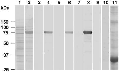
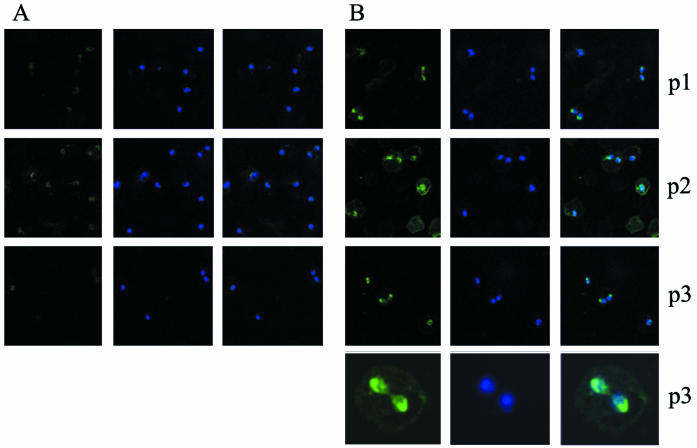
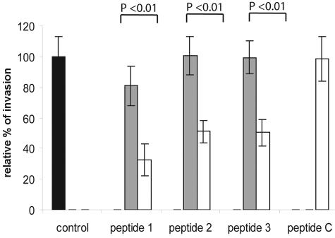
Apical membrane antigen 1 (AMA-1) is a micronemal protein secreted to the surface of merozoites of Plasmodium species and Toxoplasma gondii tachyzoites in order to fulfill an essential but noncharacterized function in host cell invasion. Here we describe cloning and characterization of a Babesia bovis AMA-1 homologue designated BbAMA-1. The overall level of similarity of BbAMA-1 to P. falciparum AMA-1 was low (18%), but characteristic features like a transmembrane domain near the C terminus, a predicted short cytoplasmic C-terminal sequence with conserved sequence properties, and an extracellular domain containing 14 conserved cysteine residues putatively involved in disulfide bridge formation are typical of AMA-1. Rabbit polyclonal antisera were raised against three synthetic peptides derived from the N-terminal region and domains II and III of the putative extracellular domain and were shown to recognize specifically recombinant BbAMA-1 expressed in Escherichia coli. Immunofluorescence microscopy showed that there was labeling of the apical half of merozoites with these antisera. Preincubation of free merozoites with all three antisera reduced the efficiency of invasion of erythrocytes by a maximum of 65%. Antisera raised against the N-terminal peptide detected a 82-kDa protein on Western blots and a 69-kDa protein in the supernatant that was harvested after in vitro invasion, suggesting that proteolytic processing and secretion take place during or shortly after invasion. A combination of two-dimensional Western blotting and metabolic labeling allowing direct identification of spots reacting with the BbAMA-1 peptide antisera together with the very low silver staining intensity of these spots indicated that very low levels of BbAMA-1 are present in Babesia merozoites.
Figures

Similar articles
-
Specific antibody to a conserved region of Babesia apical membrane antigen-1 inhibited the invasion of B. bovis into the erythrocyte.Exp Parasitol. 2013 Nov;135(3):623-8. doi: 10.1016/j.exppara.2013.09.017. Epub 2013 Sep 30. Exp Parasitol. 2013. PMID: 24090565
-
The novel protein BboRhop68 is expressed by intraerythrocytic stages of Babesia bovis.Parasitol Int. 2010 Dec;59(4):571-8. doi: 10.1016/j.parint.2010.07.008. Epub 2010 Aug 5. Parasitol Int. 2010. PMID: 20691808
-
Babesia bovis RON2 contains conserved B-cell epitopes that induce an invasion-blocking humoral immune response in immunized cattle.Parasit Vectors. 2018 Nov 3;11(1):575. doi: 10.1186/s13071-018-3164-2. Parasit Vectors. 2018. PMID: 30390674 Free PMC article.
-
Identification of candidate vaccine antigens of bovine hemoparasites Theileria parva and Babesia bovis by use of helper T cell clones.Vet Parasitol. 1995 Mar;57(1-3):189-203. doi: 10.1016/0304-4017(94)03120-l. Vet Parasitol. 1995. PMID: 7597783 Review.
-
Erythrocyte invasion by Babesia parasites: current advances in the elucidation of the molecular interactions between the protozoan ligands and host receptors in the invasion stage.Vet Parasitol. 2006 May 31;138(1-2):22-32. doi: 10.1016/j.vetpar.2006.01.037. Epub 2006 Feb 28. Vet Parasitol. 2006. PMID: 16504403 Review.
Cited by
-
Evolution of apicomplexan secretory organelles.Int J Parasitol. 2012 Nov;42(12):1071-81. doi: 10.1016/j.ijpara.2012.09.009. Epub 2012 Oct 13. Int J Parasitol. 2012. PMID: 23068912 Free PMC article. Review.
-
Babesia divergens and Plasmodium falciparum use common receptors, glycophorins A and B, to invade the human red blood cell.Infect Immun. 2005 Jan;73(1):649-51. doi: 10.1128/IAI.73.1.649-651.2005. Infect Immun. 2005. PMID: 15618210 Free PMC article.
-
Structure of AMA1 from Plasmodium falciparum reveals a clustering of polymorphisms that surround a conserved hydrophobic pocket.Proc Natl Acad Sci U S A. 2005 Sep 6;102(36):12736-41. doi: 10.1073/pnas.0501808102. Epub 2005 Aug 29. Proc Natl Acad Sci U S A. 2005. PMID: 16129835 Free PMC article.
-
Juxtamembrane shedding of Plasmodium falciparum AMA1 is sequence independent and essential, and helps evade invasion-inhibitory antibodies.PLoS Pathog. 2011 Dec;7(12):e1002448. doi: 10.1371/journal.ppat.1002448. Epub 2011 Dec 15. PLoS Pathog. 2011. PMID: 22194692 Free PMC article.
-
Identification of a highly antigenic linear B cell epitope within Plasmodium vivax apical membrane antigen 1 (AMA-1).PLoS One. 2011;6(6):e21289. doi: 10.1371/journal.pone.0021289. Epub 2011 Jun 21. PLoS One. 2011. PMID: 21713006 Free PMC article.
References
-
- Anders, R. F., P. E. Crewther, S. Edwards, M. Margetts, M. L. Matthew, B. Pollock, and D. Pye. 1998. Immunisation with recombinant AMA-1 protects mice against infection with Plasmodium chabaudi. Vaccine 16:240-247. - PubMed
-
- Anders, R. F., D. J. McColl, and R. L. Coppel. 1993. Molecular variation in Plasmodium falciparum: polymorphic antigens of asexual erythrocytic stages. Acta Trop. 53:239-253. - PubMed
-
- Bannister, L. H., J. M. Hopkins, A. R. Dluzewski, G. Margos, I. T. Williams, M. J. Blackman, C. H. Kocken, A. W. Thomas, and G. H. Mitchell. 2003. Plasmodium falciparum apical membrane antigen 1 (PfAMA-1) is translocated within micronemes along subpellicular microtubules during merozoite development. J. Cell Sci. 116:3825-3834. - PubMed
-
- Carruthers, V. B. 1999. Armed and dangerous: Toxoplasma gondii uses an arsenal of secretory proteins to infect host cells. Parasitol. Int. 48:1-10. - PubMed
-
- Cheng, Q., and A. Saul. 1994. Sequence analysis of the apical membrane antigen I (AMA-1) of Plasmodium vivax. Mol. Biochem. Parasitol. 65:183-187. - PubMed
Publication types
MeSH terms
Substances
Associated data
- Actions
LinkOut - more resources
Full Text Sources
Other Literature Sources
