. 2004 May;72(5):2956-63.
doi: 10.1128/IAI.72.5.2956-2963.2004.

Role of macrophages in host resistance to group A streptococci

Affiliations

Role of macrophages in host resistance to group A streptococci

Oliver Goldmann et al. Infect Immun. 2004 May.

Abstract

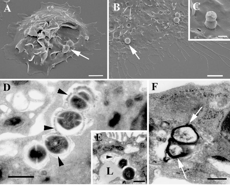
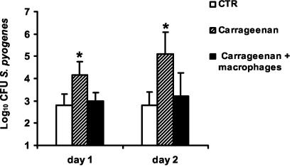
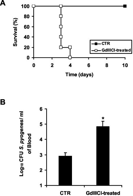
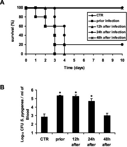
Macrophages provide the first line of defense against invading pathogens. The aim of this study was to determine the role of macrophages during infection with group A streptococci (Streptococcus pyogenes) in mice. Here, we report that resident macrophages can efficiently take up and kill S. pyogenes during in vivo infection, as demonstrated by immunofluorescence and electron microscopy, as well as colony counts. To evaluate the contribution of macrophages to the resolution of experimental infection with S. pyogenes, we compared the susceptibility of BALB/c mice rendered macrophage deficient by treatment with carrageenan with that of intact mice. The results show that depletion of macrophages enhanced the susceptibility of BALB/c mice to S. pyogenes infection, as evidenced by 100% mortality of macrophage-depleted mice compared to 90% survival of nondepleted control animals. The in vivo depletion of macrophages strongly enhanced bacterial loads in the blood and systemic organs. Resistance to S. pyogenes can be restored in macrophage-depleted mice by adoptive transfer of purified macrophages. The in vivo blocking of the macrophage phagocytic function by treatment with gadolinium III chloride also resulted in enhanced susceptibility to S. pyogenes. Interestingly, depletion of macrophages prior to or during the first 24 h of infection decreased survival dramatically; in contrast, no mortality was observed in infected nondepleted animals or mice depleted after 48 h of infection. These results emphasize the important contribution of macrophages to the early control of S. pyogenes infection.


Figures

FIG. 1.
FIG. 1.
In vivo uptake of S. pyogenes by peritoneal macrophages. Mice were intraperitoneally inoculated with either 107 (A), 5 × 10⁷ (B), or 108 (C) CFU of green-labeled S. pyogenes (solid histograms) or PBS (open histograms) and subjected to peritoneal lavage, and peritoneal macrophages were labeled with PE-conjugated anti-F4/80 antibodies. Flow cytometry analysis was performed by gating the F4/80+ population, and the association of green-labeled streptococci with macrophages was expressed as the increase in green fluorescence of macrophages (FL-1). One representative experiment out of three is shown.
FIG. 2.
FIG. 2.
Localization of S. pyogenes in peritoneal macrophages after in vivo infection. Peritoneal macrophages were isolated from mice intraperitoneally inoculated with S. pyogenes and processed for double-immunofluorescence staining. The extracellular bacteria stained green, whereas the intracellular bacteria stained red. Magnification, ×3,000. (Inset) A macrophage containing both attached (green) and internalized (red) microorganisms. Magnification, ×2,500.
FIG. 3.
FIG. 3.
Electron microscopy examination of S. pyogenes-infected peritoneal macrophages. (A to C) Scanning electron microscopy revealed streptococci (arrows) attached to macrophages (A and B) and microorganisms in the process of being internalized (C). Ultrathin sections of infected macrophages show degradation of intracellular streptococci. (D) The capsular structure (arrowheads) is being detached from the surface of the bacterium and undergoing degradation. (E) Fusion of a lysosome (L) with the phagosome. Again, degraded capsular material (arrowhead) can be identified in the phagolysosome. (F) This process leads to a complete degradation of the microorganism. Only the debris of the cell wall (arrows) can be found inside the phagolysosome. Bars, 2 (A and B), 0.5 (C), and 1 (D to F) μm.
FIG. 4.
FIG. 4.
Impaired immune defense against S. pyogenes in macrophage-depleted mice. BALB/c mice were depleted of macrophages by treatment with carrageenan and then challenged intravenously with 105 CFU of S. pyogenes. Control mice were injected with PBS. (A) Flow cytometric analysis of spleen cells isolated from control (left) or carrageenan-treated (right) animals, showing the efficiency of depletion. The dot plot in each upper right quadrant shows the F4/80 and Mac-1 profile for macrophages. (B) Survival curves of macrophage-depleted (□) and control (CTR) (▪) mice. Each group contained 10 mice. (C) Bacteremias of macrophage-depleted (solid bars) and control (open bars) mice 24 and 48 h postinoculation (n = 5 mice/group). (D) Bacterial loads in livers and spleens of macrophage-depleted (solid bars) and control (open bars) mice 48 h postinfection (n = 5 mice/group). (*, P < 0.05). One representative experiment out of four is shown. The error bars indicate standard deviations.
FIG. 5.
FIG. 5.
Restoration of anti-S. pyogenes resistance by reconstitution of carrageenan-treated mice with resident macrophages. Mice were treated with carrageenan, and one group was reconstituted with resident macrophages (solid bars) or injected with PBS (hatched bars) and then infected with S. pyogenes. Untreated mice were used as controls (CTR) (open bars). Bacteremia was determined after 24 and 48 h of bacterial inoculation. The bars represent the mean plus standard deviation of four mice per group. Statistical significance based on a comparison of carrageenan-treated and macrophage-reconstituted carrageenan-treated mice is indicated by an asterisk (P < 0.05). One representative experiment out of three is shown.
FIG. 6.
FIG. 6.
Effect of blocking macrophage phagocytic function by treatment with GdIIICl on survival of mice intravenously infected with 105 CFU of S. pyogenes (A) or on bacteremia 48 h postinfection (B). Control (CTR) mice were treated with PBS. Each bar represents the mean plus standard error of the mean of five mice per group (*, P < 0.05). One representative experiment out of four is shown.
FIG. 7.
FIG. 7.
Effect of macrophage depletion either prior to infection or 12, 24, or 48 h after intravenous inoculation on resistance against S. pyogenes. Depletion of macrophages at each time point was performed by treatment with carrageenan or PBS for control (CTR) mice. (A) Survival times of macrophage-depleted and control mice monitored over a period of 10 days. Each group contained 10 mice. (B) Bacteremias of macrophage-depleted and control mice 48 h after bacterial challenge. CFU are expressed as mean values plus standard deviations of five mice per group (*, P < 0.05). One representative experiment out of five is shown.

References

    1. Adding, L. C., G. L. Bannenberg, and L. E. Gustafsson. 2001. Basic experimental studies and clinical aspects of gadolinium salts and chelates. Cardiovasc. Drug Rev. 19:41-56. - PubMed
    1. Aderem, A. 2003. Phagocytosis and the inflammatory response. J. Infect. Dis. 187(Suppl. 2):S340-S345. - PubMed
    1. Aderem, A., and D. M. Underhill. 1999. Mechanisms of phagocytosis in macrophages. Annu. Rev. Immunol. 17:593-623. - PubMed
    1. Albanyan, E. A., and M. S. Edwards. 2000. Lectin site interaction with capsular polysaccharide mediates nonimmune phagocytosis of type III group B streptococci. Infect. Immun. 68:5794-5802. - PMC - PubMed
    1. Cone, L. A., D. R. Woodard, P. M. Schlievert, and G. S. Tomory. 1987. Clinical and bacteriologic observations of a toxic shock-like syndrome due to Streptococcus pyogenes. N. Engl. J. Med. 317:146-149. - PubMed

Publication types

LinkOut - more resources