

Differential maturation of climbing fiber innervation in cerebellar vermis
- PMID: 15102908
- PMCID: PMC6729416
- DOI: 10.1523/JNEUROSCI.5610-03.2004
Differential maturation of climbing fiber innervation in cerebellar vermis
Abstract
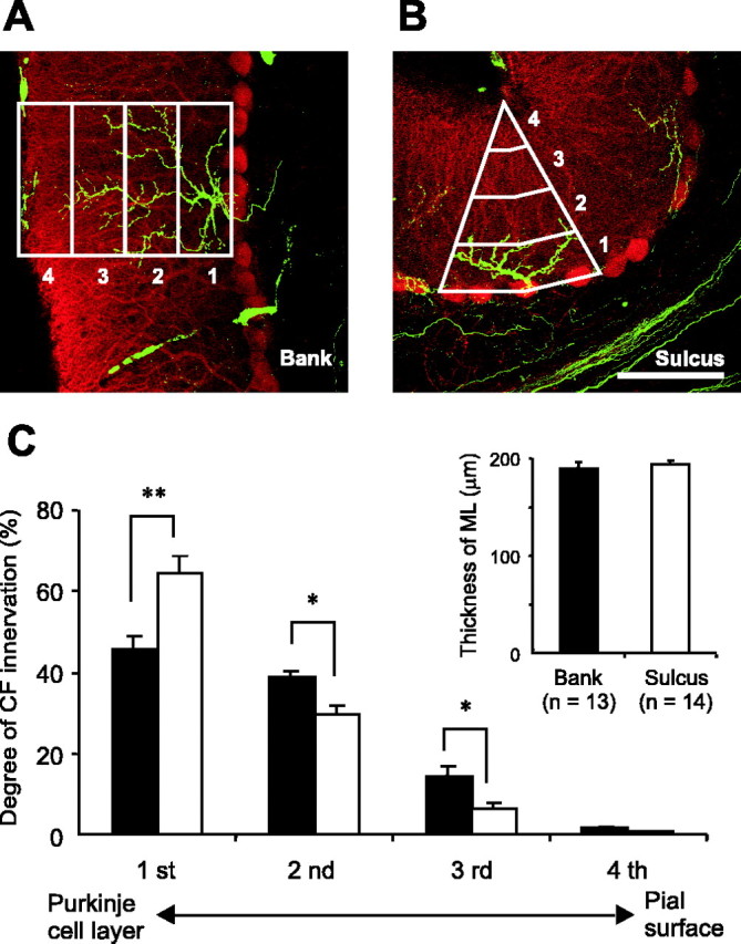
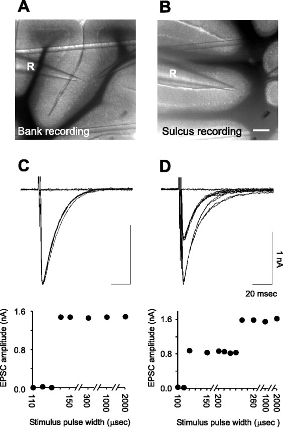
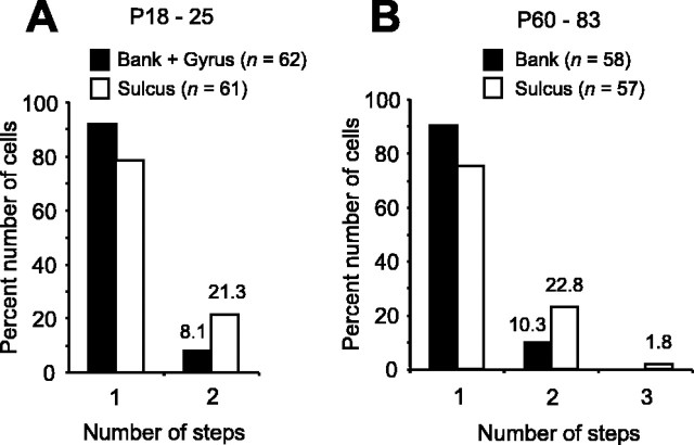
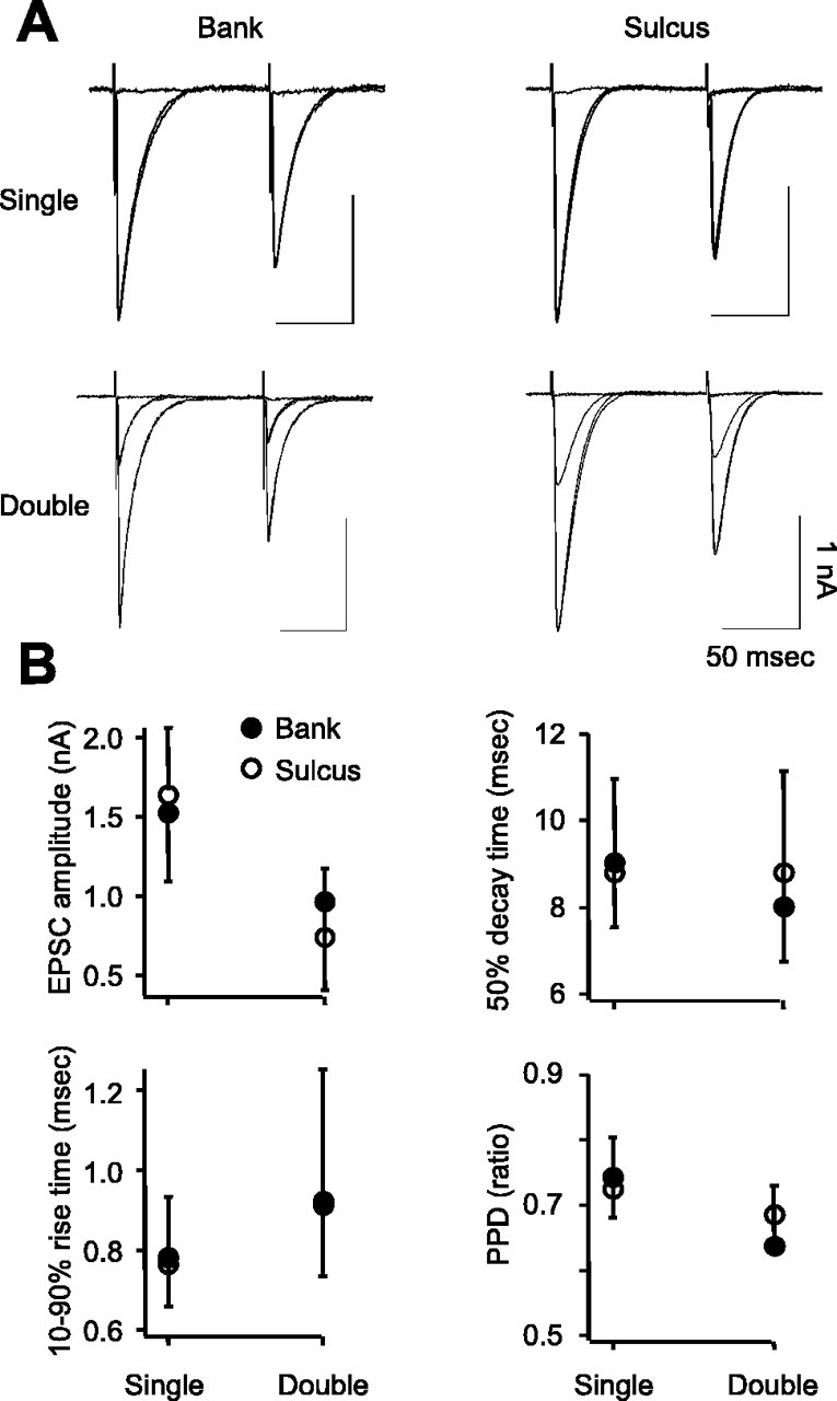
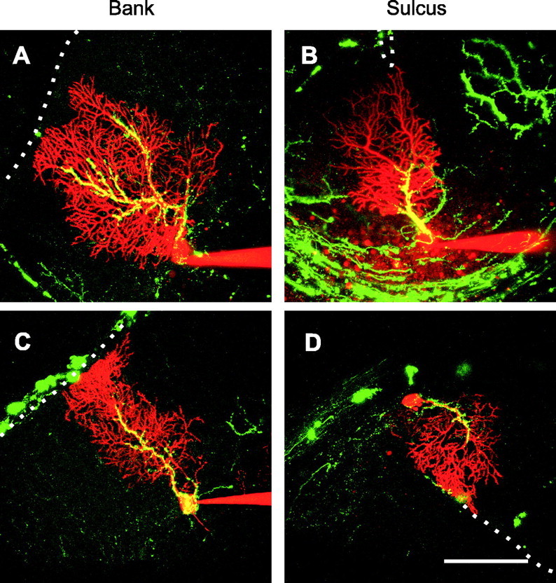
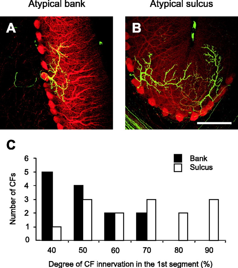
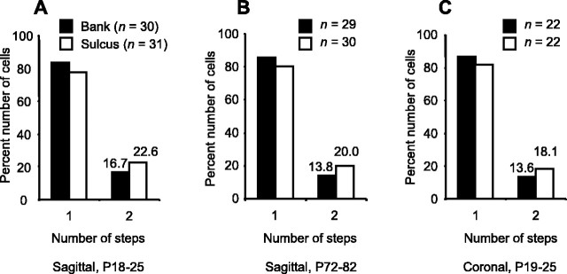
Folding of the brain surface is a general morphological adaptation to maximize surface area in a limited cranial volume. Surface folding is present not only in the neocortex but also in the cerebellar cortex. This folding creates subdivisions of the cortical surface: the sulci, the gyri, and the straight bank region, which is interposed. Is cortical folding only the solution to a surface-volume problem or does it also confer functional differences on the subdivisions that are created by this geometry? Here we have used the innervation of Purkinje cells by climbing fibers as a model system to explore potential functional differences. Purkinje cells are innervated by multiple climbing fibers at birth but undergo an activity-dependent refinement, such that by postnatal day (P) 21, most are contacted by a single climbing fiber. Using whole-cell recording from slices of cerebellar vermis derived from juvenile (P18-25) or adult (P60-83) mice, we found that significantly more Purkinje cells in the sulcus were innervated by multiple climbing fibers than in the gyrus or bank subdivisions; however, the basic properties of climbing fiber-Purkinje cell EPSCs such as kinetics, amplitude, and paired-pulse ratio were similar across cortical subdivisions. To search for a morphological correlate of differential multiple climbing fiber innervation, we labeled climbing fibers and performed reconstructions of immunofluorescent images. These revealed that, unlike the bank-gyrus subdivisions, most of the climbing fibers in the sulcus do not innervate the superficial molecular layer. These findings suggest that the subdivisions of the cerebellar cortex produced by folding may create functionally distinct entities.
Figures
Similar articles
-
A change in the pattern of activity affects the developmental regression of the Purkinje cell polyinnervation by climbing fibers in the rat cerebellum.Neuroscience. 2003;121(3):563-72. doi: 10.1016/s0306-4522(03)00556-6. Neuroscience. 2003. PMID: 14568018
-
P/Q-type Ca2+ channel alpha1A regulates synaptic competition on developing cerebellar Purkinje cells.J Neurosci. 2004 Feb 18;24(7):1734-43. doi: 10.1523/JNEUROSCI.4208-03.2004. J Neurosci. 2004. PMID: 14973254 Free PMC article.
-
Target neuron controls the integrity of afferent axon phenotype: a study on the Purkinje cell-climbing fiber system in cerebellar mutant mice.J Neurosci. 1995 Mar;15(3 Pt 1):2040-56. doi: 10.1523/JNEUROSCI.15-03-02040.1995. J Neurosci. 1995. PMID: 7891151 Free PMC article.
-
Climbing fibers mediate vestibular modulation of both "complex" and "simple spikes" in Purkinje cells.Cerebellum. 2015 Oct;14(5):597-612. doi: 10.1007/s12311-015-0725-1. Cerebellum. 2015. PMID: 26424151 Review.
-
Postnatal development and synapse elimination of climbing fiber to Purkinje cell projection in the cerebellum.Neurosci Res. 2005 Nov;53(3):221-8. doi: 10.1016/j.neures.2005.07.007. Epub 2005 Sep 1. Neurosci Res. 2005. PMID: 16139911 Review.
Cited by
-
Cerebellum morphogenesis: the foliation pattern is orchestrated by multi-cellular anchoring centers.Neural Dev. 2007 Dec 3;2:26. doi: 10.1186/1749-8104-2-26. Neural Dev. 2007. PMID: 18053187 Free PMC article.
-
TrkB is necessary for pruning at the climbing fibre-Purkinje cell synapse in the developing murine cerebellum.J Physiol. 2007 Jul 15;582(Pt 2):629-46. doi: 10.1113/jphysiol.2007.133561. Epub 2007 Apr 26. J Physiol. 2007. PMID: 17463037 Free PMC article.
-
Chronic In Vivo Imaging of Ponto-Cerebellar Mossy Fibers Reveals Morphological Stability during Whisker Sensory Manipulation in the Adult Rat.eNeuro. 2015 Oct 22;2(6):ENEURO.0075-15.2015. doi: 10.1523/ENEURO.0075-15.2015. eCollection 2015 Nov-Dec. eNeuro. 2015. PMID: 26693178 Free PMC article.
-
Axonal motility and its modulation by activity are branch-type specific in the intact adult cerebellum.Neuron. 2007 Nov 8;56(3):472-87. doi: 10.1016/j.neuron.2007.09.010. Neuron. 2007. PMID: 17988631 Free PMC article.
-
Spontaneous cluster activity in the inferior olivary nucleus in brainstem slices from postnatal mice.J Physiol. 2012 Apr 1;590(7):1547-62. doi: 10.1113/jphysiol.2011.222570. Epub 2012 Jan 16. J Physiol. 2012. PMID: 22250213 Free PMC article.
References
-
- Andjus PR, Zhu L, Cesa R, Carulli D, Strata P (2003) A change in the pattern of activity affects the developmental regression of the Purkinje cell polyinnervation by climbing fibers in the rat cerebellum. Neuroscience 121: 563-572. - PubMed
-
- Bear MF, Linden DJ (2000) The mechanisms and meaning of long-term synaptic depression. In: The synapse (Cowan WM, Südhof T, Stevens C, eds), pp 455-517. Baltimore: Johns Hopkins University.
-
- Braitenberg V, Atwood RP (1958) Morphological observations on the cerebellar cortex. J Comp Neurol 109: 1-33. - PubMed
-
- Bravin M, Rossi F, Strata P (1995) Different climbing fibres innervate separate dendritic regions of the same Purkinje cell in hypogranular cerebellum. J Comp Neurol 357: 395-407. - PubMed
Publication types
MeSH terms
Substances
Grants and funding
LinkOut - more resources
Full Text Sources
Other Literature Sources