. 2004 Apr 21;24(16):3964-73.
doi: 10.1523/JNEUROSCI.5259-03.2004.

Genetic analysis of soluble N-ethylmaleimide-sensitive factor attachment protein function in Drosophila reveals positive and negative secretory roles

Affiliations

Genetic analysis of soluble N-ethylmaleimide-sensitive factor attachment protein function in Drosophila reveals positive and negative secretory roles

Michael Babcock et al. J Neurosci. .

Abstract

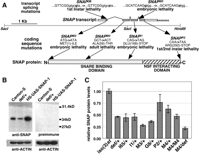
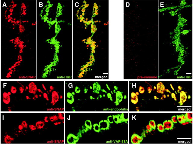
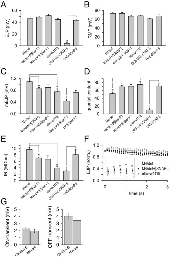
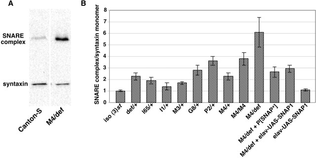
The N-ethylmaleimide-sensitive factor (NSF) and soluble NSF attachment protein (SNAP) are cytosolic factors that promote vesicle fusion with a target membrane in both the constitutive and regulated secretory pathways. NSF and SNAP are thought to function by catalyzing the disassembly of a SNAP receptor (SNARE) complex consisting of membrane proteins of the secretory vesicle and target membrane. Although studies of NSF function have provided strong support for this model, the precise biochemical role of SNAP remains controversial. To further explore the function of SNAP, we have used mutational and transgenic approaches in Drosophila to investigate the effect of altered SNAP dosage on neurotransmitter release and SNARE complex metabolism. Our results indicate that reduced SNAP activity results in diminished neurotransmitter release and accumulation of a neural SNARE complex. Increased SNAP dosage results in defective synapse formation and a variety of tissue morphological defects without detectably altering the abundance of neural SNARE complexes. The SNAP overexpression phenotypes are enhanced by mutations in other secretory components and are at least partially overcome by co-overexpression of NSF, suggesting that these phenotypes derive from a specific perturbation of the secretory pathway. Our results indicate that SNAP promotes neurotransmitter release and SNARE complex disassembly but inhibits secretion when present at high abundance relative to NSF.


Figures

Figure 1.
Figure 1.
The Drosophila SNAP mutations. A, The SNAP transcript is shown above a genomic DNA fragment that was used to rescue the SNAP mutant phenotypes. The locations of mutations likely to affect transcript splicing are shown above the SNAP transcript (capitalized sequences represent exons), and those affecting the SNAP coding sequence are shown below the transcript. B, Western blot analysis of head protein extracts from Canton-S (wild-type) flies using the anti-SNAP antiserum revealed a band of ∼35 kDa that is not detected by the preimmune serum. The size of this band is in good agreement with the expected size of SNAP protein (33 kDa). Furthermore, the intensity of this band is decreased by approximately half in lanes corresponding to a protein extract from a strain that is heterozygous for the Df(3L)rdgC-co2 deletion (def/+), which removes the SNAP gene, and significantly increased in lanes from transgenic flies overexpressing SNAP protein (HSP-GAL4/UAS-SNAP-1, designated HS-UAS-SNAP-1). C, Levels of SNAP protein present in head extracts from heterozygous SNAP mutants, SNAPM4 homozygotes, and SNAPM4 hemizygotes relative to a control [iso(3)st]. Genotype abbreviations: iso(3)st, isogenic parental chromosome; def/+, Df(3L)rdgC-co2/+; l65/+, SNAPl65/+; I1/+, SNAPI1/+; M3/+, SNAPM3/+; G8/+, SNAPG8/+; P2/+, SNAPP2/+; M4/+, SNAPM4/+; M4/M4, SNAPM4/SNAPM4; M4/def, SNAPM4/Df(3L)rdgC-co2. Error bars indicate SEM.
Figure 2.
Figure 2.
SNAP protein localizes to the presynaptic nerve terminal. Double labeling of the NMJ of larval muscles 6 and 7 with an anti-SNAP antiserum (A) and the neuronal marker anti-HRP (B) shows extensive overlap of immunoreactivity (C). Preimmune serum from rabbits immunized with SNAP protein fails to label these structures (D, E). Double-labeling experiments with anti-SNAP antiserum (F) and an antiserum to the presynaptic protein endophilin (G) reveals coincidental co-immunoreactivity (H). A single confocal section through synaptic boutons labeled with anti-SNAP antiserum (I) and antiserum to the presynaptic and postsynaptic membrane-associated protein VAP-33 (J) reveals a lack of co-localization between these markers (K). Scale bars, 5 μm.
Figure 3.
Figure 3.
Altering SNAP dosage results in attenuation of synaptic transmission. A, Average amplitude of EJPs plotted for each of six larval genotypes: SNAPM4/Df(3L)rdgC-co2 (M4/def), SNAPM4/Df(3L)rdgC-co2 P[SNAP+] (M4/def+P[SNAP+]), elavC155-GAL4;UAS-SNAP-1/+ (elav-UAS-SNAP1), elavC155-GAL4 w1118 (elav-w1118), OK6-GAL4/UAS-SNAP-5 (OK6-UAS-SNAP5), and UAS-SNAP-5/UAS-SNAP-5 (UAS-SNAP5). B, Average RMP for each genotype. C, Average amplitude of mEJPs for each genotype. D, Average quantal content for each genotype. E, Input resistance of each genotype. F, Average EJP amplitude during repetitive stimulation at 10 Hz. F, inset, Detail of the first five EJP amplitudes of the 10 Hz train. G, Average absolute amplitudes of the ON and OFF transients plotted for SNAPM4/Df(3L)rdgC-co2 (M4/def) and wild-type controls (Canton-S). Error bars indicate SEM. *,#Significant differences at p < 0.05; *one-way ANOVA followed by a Student-Newman-Keuls pair wise multiple-comparison test; #unpaired (2-tailed) t test.
Figure 4.
Figure 4.
SNARE complex abundance is increased in SNAP mutants. Protein extracts from adult heads were subjected to Western blot analysis with an mAb that recognizes the SNARE protein syntaxin. A, Representative Western blot depicting the SNARE complex and syntaxin monomer in a wild-type control sample (Canton-S) and animals bearing the SNAPM4 mutation in trans to the Df(3L)rdgC-co2 deletion (M4/def). Note the increase in SNARE complex abundance and decrease in syntaxin monomer abundance in the hemizygous SNAPM4 mutant relative to Canton-S. B, The ratio of SNARE complex abundance to syntaxin monomer abundance is plotted relative to iso(3)st (for details, see Materials and Methods). Genotype abbreviations are as defined in Figures 1 and 3 but also include the following: M4/def + elav-UAS-SNAP1, elavC155;UAS-SNAP-1/+;SNAPM4/Df(3L)rdgC-co2; elav-UAS-SNAP1, elavC155;UAS-SNAP-1/+. All of the genotypes shown manifest significant differences from the iso(3)st control with the exception of I1/+ and elav-UAS-SNAP-1 by unpaired (2-tailed) t tests (p < 0.05). Additionally, M4/def + P[SNAP+] and M4/def + elav-UAS-SNAP1 manifest significant differences from the M4/def alone using the above test. Error bars indicate SEM.
Figure 5.
Figure 5.
SNAP overexpression disrupts neuromuscular junction morphology. The larval NMJs of muscles 6 and 7 were subject to immunohistochemistry using antisera to the axonal filament protein futch (green) and the synaptic vesicle protein synaptotagmin (red). NMJ morphology in SNAPM4 hemizygotes (B) is similar to that in control larvae (A). Use of the Ok6-GAL4 line to drive overexpression of SNAP protein from the UAS-SNAP-5 transgene results in a dramatically reduced synaptic bouton number (D) relative to a control (C). Scale bars: A, B,10 μm; C, D, 50 μm.
Figure 6.
Figure 6.
Synaptic vesicle size, distribution, and number are unaltered in SNAPM4 mutants. A, B, Representative electron micrographs of the larval NMJ at muscle 6 and 7 segment 3 from Canton-S control larvae (A) and SNAPM4/Df(3L)rdgC-co2 hemizygotes (M4/def; B). C, D, Higher-magnification images of synaptic vesicles surrounding the t-bar structures located at sites of neurotransmitter release in control larvae (C) and larvae hemizygous for the SNAPM4 mutation (D). The average diameter of synaptic vesicles in SNAPM4 hemizygotes is not significantly different from that of Canton-S using unpaired (2-tailed) t tests (E). The distribution of synaptic vesicles within 200 nm of release sites (F) is grossly similar in SNAPM4 hemizygotes and Canton-S. The total numbers of synaptic vesicles within 200 nm of release sites in Canton-S (33.0 ± 1.8) and SNAPM4 hemizygotes (35.4 ± 1.8) are also within experimental error of one another. Scale bars: A, B, 100 nm; C, D, 10 nm.
Figure 7.
Figure 7.
SNAP overexpression phenotypes are modified by altered dosage of other secretory components. Pan-neuronal expression of the UAS-SNAP-1 transgene using the elavC155-GAL4 driver results in a wing inflation defect (A), and co-expression of either dNSF1 or dNSF2 completely suppresses this phenotype (B). GMR-GAL4-driven expression of UAS-SNAP-1 produces a mildly rough eye showing patches of necrosis, mild furrowing at ommatidial borders, and progressive degeneration (D) with respect to wild type (C). GMR-GAL4-driven expression of the stronger UAS-SNAP-5 results in near total ablation of the eye (E). The presence of a single copy of the dNSF2l55 mutation (F) or the syxΔ229 mutation (G) in the GMR-GAL4 UAS-SNAP-1 background resulted in enhancement of the phenotypes resulting from ectopic expression of SNAP in the eye, including increased necrosis around the perimeter of the compound eye and more dramatic furrowing at ommatidial junctions (bottom panels). Phenotypes associated with GMR-GAL4-driven expression of UAS-SNAP-1 and UAS-SNAP-5 are suppressed by co-expression of either dNSF1 or dNSF2 (H-K). Genotype abbreviations include the following: Elav-UAS-SNAP1, elavC155-GAL4/+;UAS-SNAP-1/+; Elav-UAS-SNAP1 UAS-NSF1, elavC155-GAL4/+;UAS-SNAP-1/UAS-NSF1(73C); Elav-UAS-SNAP1 UAS-NSF2, elavC155-GAL4/+;UAS-SNAP-1/UAS-NSF2(23A); GMR-UAS-SNAP1, GMR-GAL4/UAS-SNAP-1; GMR-UAS-SNAP5, GMR-GAL4/UAS-SNAP-5; GMR-UAS-SNAP1 nsf2l-55, GMR-GAL4 UAS-SNAP-1/nsf2l-55; GMR-UAS-SNAP1 syx1AΔ229, GMR-GAL4 UAS-SNAP-1/syx1AΔ229; GMR-UAS-SNAP1 UAS-NSF1, GMR-GAL4 UAS-SNAP-1/UAS-dNSF1(73C); GMR-UAS-SNAP1 UAS-NSF2, GMR-GAL4 UAS-SNAP-1/UAS-dNSF2(23A); GMR-UAS-SNAP5 UAS-NSF1, GMR-GAL4/UAS-dNSF1(73C) UAS-SNAP-5; GMR-UAS-SNAP5 UAS-NSF2, GMR-GAL4 UAS-dNSF2(23A)/UAS-SNAP-5.

References

    1. Aberle H, Haghighi AP, Fetter RD, McCabe BD, Magalhaes TR, Goodman CS (2002) Wishful thinking encodes a BMP type II receptor that regulates synaptic growth in Drosophila Neuron 33: 545-558. - PubMed
    1. Barnard RJ, Morgan A, Burgoyne RD (1997) Stimulation of NSF ATPase activity by alpha-SNAP is required for SNARE complex disassembly and exocytosis. J Cell Biol 139: 875-883. - PMC - PubMed
    1. Bellen HJ, Budnik V (2000) The neuromuscular junction. In: Drosophila protocols (Sullivan W, Hawley RS, Ashburner M, eds), pp 175-199. Cold Spring Harbor, NY: Cold Spring Harbor Laboratory.
    1. Budnik V, Zhong Y, Wu CF (1990) Morphological plasticity of motor axons in Drosophila mutants with altered excitability. J Neurosci 10: 3754-3768. - PMC - PubMed
    1. Burg MG, Sarthy PV, Koliantz G, Pak WL (1993) Genetic and molecular identification of a Drosophila histidine decarboxylase gene required in photoreceptor transmitter synthesis. EMBO J 12: 911-919. - PMC - PubMed

Publication types

MeSH terms

Substances

LinkOut - more resources