. 2004 Apr 21;24(16):3974-84.
doi: 10.1523/JNEUROSCI.3720-03.2004.

Specific Na+ sensors are functionally expressed in a neuronal population of the median preoptic nucleus of the rat

Affiliations

Specific Na+ sensors are functionally expressed in a neuronal population of the median preoptic nucleus of the rat

Magali Grob et al. J Neurosci. .

Abstract

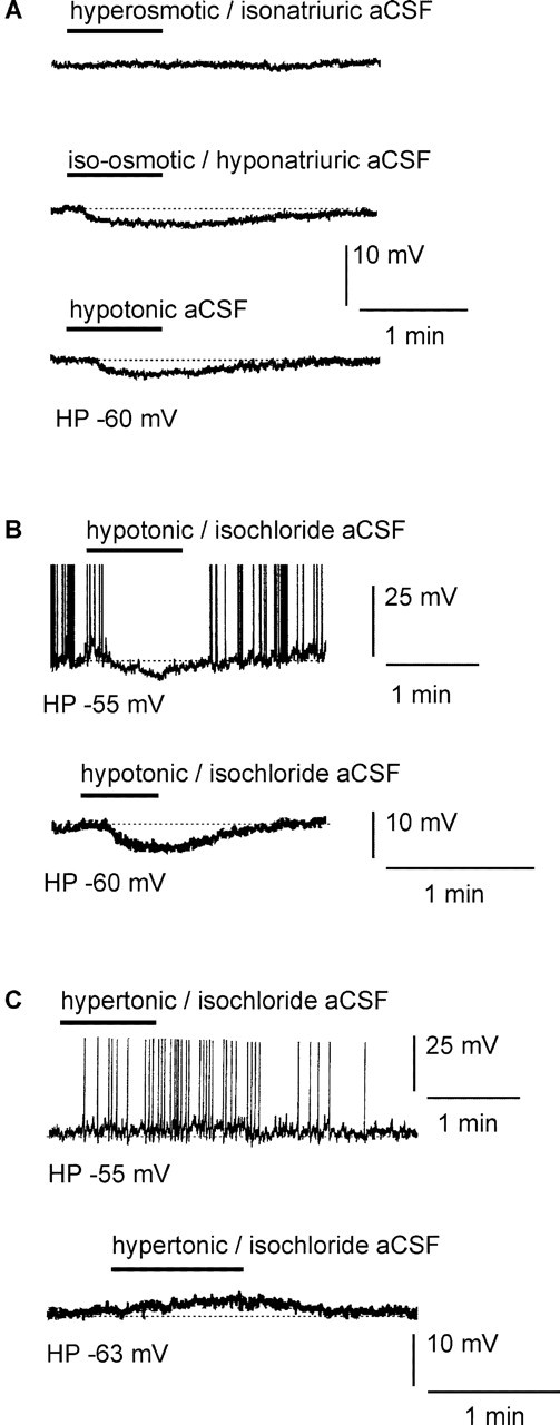
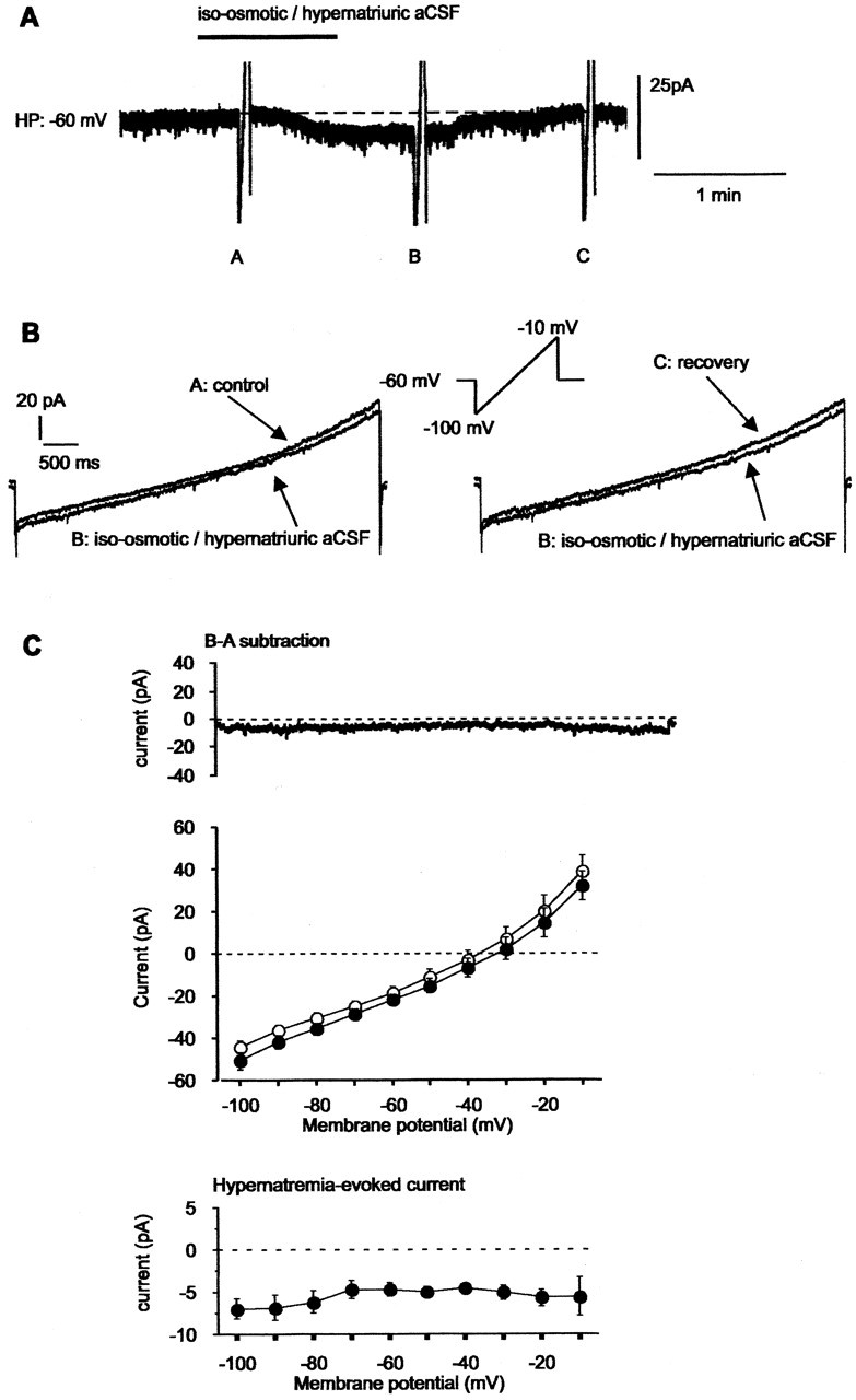
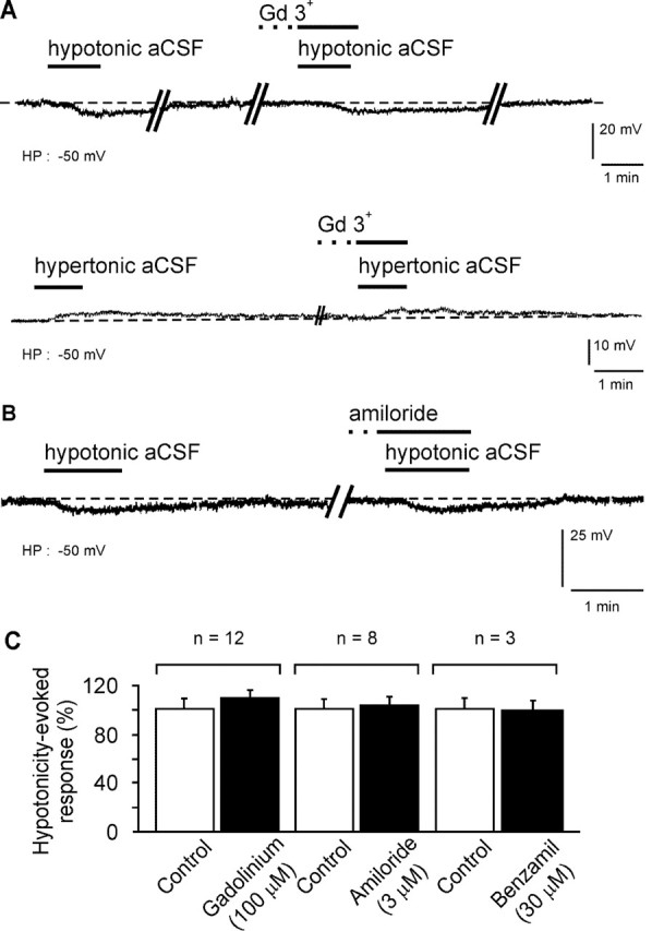
Whole-cell patch-clamp recordings were performed on acute brain slices of male rats to investigate the ability of the neurons of the median preoptic nucleus (MnPO) to detect fluctuation in extracellular osmolarity and sodium concentration ([Na+]out). Local application of hypotonic and hypertonic artificial CSF hyperpolarized and depolarized the neurons, respectively. Similar responses obtained under synaptic isolation (0.5 microM TTX) highlighted the intrinsic ability of the MnPO neurons to detect changes in extracellular osmolarity and [Na+]out. Manipulating extracellular osmolarity, [Na+]out, and [Cl-]out showed in an independent manner that the MnPO neurons responded to a change in [Na+]out exclusively. The specific Na+ response was voltage insensitive and depended on the driving force for Na+ ions, indicating that a sustained background Na+ permeability controlled the membrane potential of the MnPO neurons. This specific response was not reduced by Gd3+, amiloride, or benzamil, ruling out the participation of mechanosensitive cationic channels, specific epithelial Na+ channels, and Phe-Met-Arg-Phe-gated Na+ channels, respectively. Combination of in situ hybridization, using a riboprobe directed against the atypical Na+ channel (Na(X)), and immunohistochemistry, using an antibody against neuron-specific nuclei protein, revealed that a substantial population of MnPO neurons expressed the Na(X) channel, which was characterized recently as a concentration-sensitive Na+ channel. This study shows that a neuronal population of the MnPO acts as functional Na+ sensors and that the Na(X) channel might represent the molecular basis for the extracellular sodium level sensing in these neurons.


Figures

Figure 7.
Figure 7.
NaX channel is expressed in the median preoptic nucleus and subfornical organ. The line drawings, adapted from the brain atlas of Swanson (1992), represent coronal sections of rat brain through the subfornical organ (A, left) and the median preoptic nucleus (B, left). The autoradiograms illustrate the distribution of NaX mRNA in the subfornical organ (A, right) and in the ventral portion of median preoptic nucleus (B, right). Note that in situ hybridization performed with the sense probe did not reveal any signal in both the SFO and MnPO (A, B, middle, white arrows). These pictures were taken from autoradiogram of 30 μm coronal sections hybridized with 35S-labeled NaX riboprobe through corresponding sections. 3rd V, Third ventricle; LV, lateral ventricle.
Figure 1.
Figure 1.
Intrinsic osmosensitivity is a unique property of the MnPO neurons. A, Depolarization of a ventral MnPO neuron with current injection triggered a regular spiking activity. Local and transient (1min) application of hypotonic aCSF (270 mOsm/l; 100 mm NaCl) over the vMnPO region hyperpolarized the neuron with a concomitant abolition of the spike discharges (top trace). The same neuron was held below the spike threshold, and local application of hypertonic aCSF (330 mOsm/l; 170 mm NaCl) depolarized the neuron that discharged a prolonged burst of spikes (bottom trace). Details of the spiking activity was illustrated before (*) and after (**) application of the modified aCSF. B, TTX (0.5 μm) was bath applied to block synaptic inputs onto the recorded vMnPO neuron. Under this steady-state synaptic blockage, transient and local application of a hypotonic aCSF hyperpolarized the neuron (top trace). In contrast, local application of a hypertonic aCSF induced a depolarization (bottom trace). C, Local application of hypertonic aCSF did not alter the membrane potential of this vMnPO neuron, which was considered as a non-osmoresponsive neuron. D, Local application of hypotonic aCSF did not change the membrane potential of the neurons located in a region adjacent to the MnPO, the median septum. Note that spike amplitude has been truncated in the traces presented here. These results indicate that detection of the extracellular tonicity involved intrinsic properties of a specific neuronal population of the vMnPO. HP, Holding potential.
Figure 2.
Figure 2.
Ventral MnPO neurons detect changes in extracellular Na+ level but not osmolarity. A, Local application of a hyperosmotic-isonatriuric stimulus (330 mOsm/l; 150 mm NaCl) had no apparent effect on the excitability of the vMnPO neuron (top trace). In contrast, local application of an iso-osmotic-hyponatriuric aCSF (300 mOsm/l; 100 mm NaCl) hyperpolarized the vMnPO neuron (middle trace). The amplitude of the hyperpolarization was similar to the one obtained with a hypotonic aCSF (270 mOsm/l; 100 mm NaCl; bottom trace). B, Traces obtained from two different vMnPO neurons. The top trace was obtained with slight injection of depolarizing current to make the neuron fire (holding potential, -55 mV). The bottom trace was obtained from a neuron maintained around its resting potential (holding potential, -60 mV). In these neurons, transient and local application of hypotonic-isochloride aCSF (270 mOsm/l; 100 mm NaCl and 50mm choline chloride) drove the membrane potential to hyperpolarization. C, Two neurons maintained at different membrane potential[holding potential, -55 mV (top trace); holding potential, -63 mV (bottom trace)]. Local application of hypertonic-isochloride aCSF (330 mOsm/l; 150mm NaCl and 20 mm Na+-gluconate) onto these neurons triggered depolarization of the cells. Spike amplitude has been truncated in all of the panels presented. These results show that neurons of the vMnPO did respond to a change in extracellular [Na+] and not to an alteration of the extracellular osmolarity or [Cl-]. HP, Holding potential.
Figure 3.
Figure 3.
The specific response to a natriuric challenge depended on the driving force for Na+ ions and was not associated with a change in input resistance. A, In the same vMnPO neuron, local application of iso-osmotic-hyponatriuric aCSF with a reduction of 8, 20, and 50 mm in the [Na+]out compared with regular aCSF (150 mm) induced a cellular response of graded amplitude. B, Local application of iso-osmotic-hyponatriuric aCSF (300 mOsm/l; 100 mm NaCl) induced membrane hyperpolarization that was not accompanied with a change in input resistance. Input resistance was tested with 800 msec hyperpolarizing current steps (-8 pA) tested before and during the cellular response. Note that positive current was transiently injected to compensate for the hypotremia-induced hyperpolarization. Typical representation of the passive membrane response to the current step is an average of five consecutive traces. C, Same neuron as in B. Local application of hypernatriuric aCSF (330 mOsm/l; 170 mm NaCl) induced a depolarization that was not accompanied by a change in input resistance. Input resistance was assessed with identical current steps as those depicted in B. Negative current was transiently injected to the cell to compensate for the hypernatremia-induced depolarization during evaluation of the input resistance. HP, Holding potential.
Figure 4.
Figure 4.
Transient application of iso-osmotic-hyponatriuric aCSF on vMnPO neurons triggered a voltage-independent outward current. A, A vMnPO neuron was recorded under the voltage-clamp mode and maintained at a potential of -60 mV. Local application of iso-osmotic-hyponatriuric aCSF (300 mOsm/l; 100 mm NaCl) triggered an outward current. B, Three I-V relationships from -100 to -10 mV (16 mV/sec) were elicited under control (labelA), iso-osmotic-hyponatriuric aCSF (labelB), and back to iso-osmotic-isonatriuric aCSF (label C). The specific current (Δ current) evoked by hyponatremia was isolated by digital subtraction of the I-V relationships recorded in A and B. Note the absence of voltage dependency over the range of potentials tested. TTX (0.5 μm) and TEA-Cl (20 mm) were present in isotonic aCSF, as well as in iso-osmotic-hyponatriuric aCSF, to block voltage-gated Na+ and K+ currents. For these experiments, extracellular osmolarity (300 mOsm/l) was achieved with or without adding mannitol to aCSF (iso-osmotic-hyponatriuric aCSF and control, respectively). C, Mean ramp current obtained from the protocol described in B (n = 6 MnPO neurons), showing a parallel shift of the current in the outward direction during transient application of iso-osmotic-hyponatriuric aCSF (top). Mean control current is represented by open circles, and mean current recorded during application of modified aCSF is represented by filled circles. Digital subtraction of the traces shown in the top panel resolved the I-V relationship of the mean current evoked by application of iso-osmotic-hyponatriuric aCSF (bottom). HP, Holding potential.
Figure 5.
Figure 5.
Transient application of iso-osmotic-hypernatriuric aCSF on vMnPO neurons triggered a voltage-independent inward current. A, A vMnPO neuron was maintained at a membrane potential of -60 mV. Local application of iso-osmotic-hypernatriuric aCSF (300 mOsm/l; 150 mm NaCl) compared with control solution (300 mOsm/l; 100 mm NaCl) triggered an inward current. B, I-V relationships from -100 to -10 mV (16 mV/sec) were elicited before (label A), during (label B), and after (label C) the application of iso-osmotic-hypernatriuric aCSF. The specific current (Δ current) evoked by hypernatremia was isolated by digital subtraction of the I-V relationships recorded in A and B. TTX (0.5 μm) and TEA-Cl (20 mm) were present in control aCSF, as well as in iso-osmotic-hypernatriuric aCSF to block voltage-gated Na+ and K+ currents. For these experiments, extracellular osmolarity (300 mOsm/l) was achieved by adding mannitol to the control aCSF and by removing glucose from the iso-osmotic-hypernatriuric aCSF. C, Mean ramp current obtained from the protocol described in B and obtained from the six MnPO neurons illustrated in Figure 4C. Transient application of iso-osmotic-hypernatriuric aCSF evoked an inward shift of the current (top). Mean current recorded under control is represented by open circles, and mean current recorded during application of modified aCSF is represented by filled circles. Digital subtraction of the traces shown in the top panel resolved the I-V relationship of the mean current evoked by application of iso-osmotic-hypernatriuric aCSF (bottom). HP, Holding potential.
Figure 6.
Figure 6.
The pharmacology of the specific Na+ response ruled out the involvement of mechanosensitive cationic channels, as well as amiloride-sensitive Na+ channels. A, Typical response of a neuron of the vMnPO to local application of hypotonic (270 mOsm/l; 100 mm NaCl) and hypertonic (330 mOsm/l; 170 mm NaCl) aCSF, recorded in the presence of TTX in the bath (0.5 μm). Note that bath application of Gd3+ before the hypotonic and hypertonic stimuli did not reduce the amplitude of the hyperpolarization and depolarization of the vMnPO neurons, respectively. B, In a second set of vMnPO neurons, steady-state application of amiloride, a blocker of specific brain Na+ channels, remained ineffective for blocking the hyperpolarization induced by local application of a hypotonic aCSF. C, Histograms summarizing the pharmacology of the Na+ response mediated by local application of a hypotonic stimulus. The action of gadolinium, amiloride, and benzamil was tested on 12, 8, and 3 neurons, respectively. HP, Holding potential.
Figure 8.
Figure 8.
Neuronal populations of the median preoptic nucleus express NaX. A-H, Bright-field photomicrographs obtained from two different frontal sections of the brain that include the MnPO. B, F, A general view of the selected area (MnPO), together with adjacent control regions [lateral septum and bed nucleus of the stria terminalis (BnST)]. A,D,E, Photomicrographs at different magnifications showing the distribution of both the immunoreactivity for neuron-specific nuclei protein and NaX mRNA at the level of the MnPO. The arrows in A and E illustrate double-labeled cells that are in the same focal plan for the Nax channel mRNA (black silver grains) and neuron-specific nuclei protein (immunoreactivity in brown). C, G, H, Photomicrographs of control regions taken at different magnifications. Note the absence of double-labeled neurons in surrounding nuclei, as shown for the septum (C) and the bed nucleus of the stria terminalis (H). Scale bars: A, C, E, H, 20 μm; B, F, 400 μm; D, G, 50 μm. 3V, Third ventricle; LV, lateral ventricle; ac, anterior commissure.

Similar articles

Cited by

References

    1. Akopian AN, Souslova V, Sivilotti L, Wood JN (1997) Structure and distribution of a broadly expressed atypical sodium channel. FEBS Lett 400: 183-187. - PubMed
    1. Aradachi H, Honda K, Negoro H, Kubota T (1996) Median preoptic neurones projecting to the supraoptic nucleus are sensitive to haemodynamic changes as well as to rise in plasma osmolality in rats. J Neuroendocrinol 8: 35-43. - PubMed
    1. Black JA, Yokoyama S, Waxman SG, Oh Y, Zur KB, Sontheimer H, Higashida H, Ransom BR (1994) Sodium channel mRNAs in cultured spinal cord astrocytes: in situ hybridization in identified cell types. Brain Res Mol Brain Res 23: 235-245. - PubMed
    1. Bourque CW, Oliet SHR, Richard D (1994) Osmoreceptors, osmoreception and osmoregulation. Front Neuroendocrinol 15: 231-274. - PubMed
    1. Cox PS, Denton DA, Mouw DR, Tarjan E (1987) Natriuresis induced by localized perfusion within the third cerebral ventricle of sheep. Am J Physiol 252: R1-R6. - PubMed

Publication types

MeSH terms

LinkOut - more resources