

Dual neuroprotective signaling mediated by downregulating two distinct phosphatase activities of PTEN
- PMID: 15102920
- PMCID: PMC6729419
- DOI: 10.1523/JNEUROSCI.5449-03.2004
Dual neuroprotective signaling mediated by downregulating two distinct phosphatase activities of PTEN
Abstract
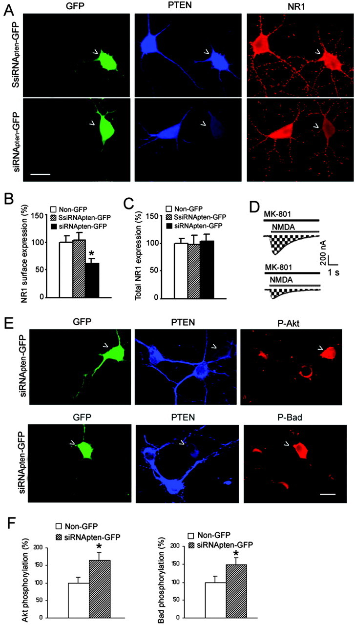
The tumor suppressor PTEN (phosphatase and tensin homolog deleted on chromosome 10) is a lipid and protein phosphatase. We report here that PTEN physically associates with the NR1 and NR2B subunits of NMDA receptors (NMDARs) in rat hippocampus. Downregulating the protein expression of PTEN inhibits the function of extrasynaptic NMDARs and decreases NMDAR surface expression, suggesting a crucial role for endogenous PTEN in the modulation of NMDAR-mediated neuronal function. Reducing PTEN expression also enhances Akt/Bad phosphorylation in hippocampal neurons. Importantly, suppressing lipid and protein phosphatase activity of PTEN, respectively, activates Akt and inhibits extrasynaptic NMDAR activity and thereby protects against ischemic neuronal death in vitro and in vivo. Thus, our study reveals a dual neuroprotective mechanism by which Akt/Bad and extrasynaptic NMDARs are regulated via downregulation of two distinct PTEN phosphatase activities and present the possibility of PTEN as a potential therapeutic target for stroke treatment.
Figures


Similar articles
-
Phosphorylation of PTEN and Akt in astrocytes of the rat hippocampus following transient forebrain ischemia.Cell Tissue Res. 2005 Mar;319(3):359-66. doi: 10.1007/s00441-004-1033-0. Epub 2005 Jan 27. Cell Tissue Res. 2005. PMID: 15726426
-
Phospho-regulation of synaptic and extrasynaptic N-methyl-d-aspartate receptors in adult hippocampal slices.Neuroscience. 2009 Feb 18;158(4):1446-59. doi: 10.1016/j.neuroscience.2008.11.006. Epub 2008 Nov 8. Neuroscience. 2009. PMID: 19041929
-
Anti-apoptotic action of (2S,3S,4R)-N"-cyano-N-(6-amino-3,4-dihydro-3-hydroxy-2-methyl-2-dimethoxymethyl-2H-benzopyran-4-yl)-N'-benzylguanidine (KR-31378) by suppression of the phosphatase and tensin homolog deleted from chromosome 10 phosphorylation and increased phosphorylation of casein kinase2/Akt/ cyclic AMP response element binding protein via maxi-K channel opening in neuronal cells.Eur J Pharmacol. 2004 Aug 30;497(3):267-77. doi: 10.1016/j.ejphar.2004.06.058. Eur J Pharmacol. 2004. PMID: 15336944
-
Pten signaling in gliomas.Neuro Oncol. 2002 Jul;4(3):196-211. Neuro Oncol. 2002. PMID: 12084351 Free PMC article. Review.
-
Biological role of phosphatase PTEN in cancer and tissue injury healing.Front Biosci. 2002 May 1;7:e245-51. doi: 10.2741/tsugawa. Front Biosci. 2002. PMID: 11991859 Review.
Cited by
-
Glycine confers neuroprotection through microRNA-301a/PTEN signaling.Mol Brain. 2016 May 26;9(1):59. doi: 10.1186/s13041-016-0241-3. Mol Brain. 2016. PMID: 27230112 Free PMC article.
-
Role of NMDA Receptor-Mediated Glutamatergic Signaling in Chronic and Acute Neuropathologies.Neural Plast. 2016;2016:2701526. doi: 10.1155/2016/2701526. Epub 2016 Aug 18. Neural Plast. 2016. PMID: 27630777 Free PMC article. Review.
-
The Need for SMN-Independent Treatments of Spinal Muscular Atrophy (SMA) to Complement SMN-Enhancing Drugs.Front Neurol. 2020 Feb 3;11:45. doi: 10.3389/fneur.2020.00045. eCollection 2020. Front Neurol. 2020. PMID: 32117013 Free PMC article. Review.
-
Exosomal miR-9 Released from HIV Tat Stimulated Astrocytes Mediates Microglial Migration.J Neuroimmune Pharmacol. 2018 Sep;13(3):330-344. doi: 10.1007/s11481-018-9779-4. Epub 2018 Mar 1. J Neuroimmune Pharmacol. 2018. PMID: 29497921 Free PMC article.
-
L-Serine, an Endogenous Amino Acid, Is a Potential Neuroprotective Agent for Neurological Disease and Injury.Front Mol Neurosci. 2021 Sep 6;14:726665. doi: 10.3389/fnmol.2021.726665. eCollection 2021. Front Mol Neurosci. 2021. PMID: 34552468 Free PMC article. Review.
References
-
- Backman SA, Stambolic V, Suzuki A, Haight J, Elia A, Pretorius J, Tsao MS, Shannon P, Bolon B, Ivy GO, Mak TW (2001) Deletion of Pten in mouse brain causes seizures, ataxia and defects in soma size resembling Lhermitte-Duclos disease. Nat Genet 29: 396-403. - PubMed
-
- Barria A, Malinow R (2002) Subunit-specific NMDA receptor trafficking to synapses. Neuron 35: 345-353. - PubMed
-
- Beattie EC, Carroll RC, Yu X, Morishita W, Yasuda H, von Zastrow M, Malenka RC (2000) Regulation of AMPA receptor endocytosis by a signaling mechanism shared with LTD. Nat Neurosci 3: 1291-1300. - PubMed
-
- Brewer GJ, Torricelli JR, Evege EK, Price PJ (1993) Optimized survival of hippocampal neurons in B27-supplemented Neurobasal, a new serum-free medium combination. J Neurosci Res 35: 567-576. - PubMed
-
- Brummelkamp TR, Bernards R, Agami R (2002) A system for stable expression of short interfering RNAs in mammalian cells. Science 296: 550-553. - PubMed
Publication types
MeSH terms
Substances
LinkOut - more resources
Full Text Sources
Other Literature Sources
Medical
Molecular Biology Databases
Research Materials