Comparative Study
. 2004 May 5;23(9):1922-33.
doi: 10.1038/sj.emboj.7600203. Epub 2004 Apr 22.

Svp1p defines a family of phosphatidylinositol 3,5-bisphosphate effectors

Affiliations
Comparative Study

Svp1p defines a family of phosphatidylinositol 3,5-bisphosphate effectors

Stephen K Dove et al. EMBO J. .

Abstract

Phosphatidylinositol 3,5-bisphosphate (PtdIns(3,5)P2), made by Fab1p, is essential for vesicle recycling from vacuole/lysosomal compartments and for protein sorting into multivesicular bodies. To isolate PtdIns(3,5)P2 effectors, we identified Saccharomyces cerevisiae mutants that display fab1delta-like vacuole enlargement, one of which lacked the SVP1/YFR021w/ATG18 gene. Expressed Svp1p displays PtdIns(3,5)P2 binding of exquisite specificity, GFP-Svp1p localises to the vacuole membrane in a Fab1p-dependent manner, and svp1delta cells fail to recycle a marker protein from the vacuole to the Golgi. Cells lacking Svp1p accumulate abnormally large amounts of PtdIns(3,5)P2. These observations identify Svp1p as a PtdIns(3,5)P2 effector required for PtdIns(3,5)P2-dependent membrane recycling from the vacuole. Other Svp1p-related proteins, including human and Drosophila homologues, bind PtdIns(3,5)P2 similarly. Svp1p and related proteins almost certainly fold as beta-propellers, and the PtdIns(3,5)P2-binding site is on the beta-propeller. It is likely that many of the Svp1p-related proteins that are ubiquitous throughout the eukaryotes are PtdIns(3,5)P2 effectors. Svp1p is not involved in the contributions of FAB1/PtdIns(3,5)P2 to MVB sorting or to vacuole acidification and so additional PtdIns(3,5)P2 effectors must exist.


Figures

Figure 1
Figure 1
The svp1Δ phenotype involves vacuole enlargement, and GFP-Svp1p localisation at the vacuole membrane is FAB1-dependent. (A) Differential interference contrast images and FM4-64 staining of wild-type, svp1Δ and fab1Δ cells, demonstrating the greatly enlarged vacuoles of svp1Δ cells. (B) Svp1p expression corrects the svp1Δ vacuolar defect (upper images, taken during methionine-repressed low-level Svp1p expression), but Svp1p overexpression induces cell vacuolation (lower images, during de-repressed expression following methionine removal). (C) When GFP-Svp1p was expressed in svp1Δ cells, it associated mainly with the vacuole membrane and large punctate structures. Little or none of the GFP-Svp1p was associated with the vacuole membrane in fab1Δ cells.
Figure 2
Figure 2
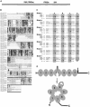
Sequence analysis and structural modelling of Svp1p and Svp1p-like proteins. (A) YFR021w/SVP1 is on the right arm of chromosome VI, adjacent to FAB1. (B) ClustalW alignment of some eukaryotic Svp1-like proteins. Light-grey bars identify sequence within the putative β-propeller. The dark-grey bar denotes the B/C insert in blade 4. The black arrow shows the deduced site of trypsin cleavage in Svp1p. The black bar indicates the C-terminal domain outside the β-propeller. (C) Threaded alignment of Svp1p, hSvp1a and Hsv2p with the β-propeller of transducin-β (Sondek et al, 1996). Assignment of blades and β-strands is based on transducin-β. Each sequence was submitted to the 3D-PSSM server (Kelley et al, 2000), and gave a significant (>80% certainty) score for alignment with transducin-β. Alignments were slightly adjusted in blades 3, 4 and 7, to maintain consistency within the Svp1p family ClustalW alignments. (D) A linear depiction of the Svp1p domain structure and a cartoon of its probable folded structure. Light ovals represent WD-40 blades, and the black arrow the point of trypsin attack.
Figure 3
Figure 3
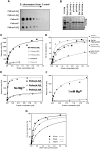
Svp1p and related proteins bind PtdIns(3,5)P2 with high affinity and selectivity. (A) A dot-blot assay indicates that GST-Svp1p binds both PtdIns3P and PtdIns(3,5)P2. (B) GST-Svp1p bound to PtdIns(3,5)P2-derivatised Affigel beads is selectively displaced by exogenous PtdIns(3,5)P2 and not by other PtdinsP2 isomers or by PtdInsPs. (C) Monomeric Svp1p binds to a DOPC layer ‘doped' with 3 mol% PtdIns(3,5)P2 but not with other phosphoinositides, as detected by Biacore analysis. (D) The affinity of monomeric Svp1p binding to PtdIns(3,5)P2-‘doped' lipids is similar to phosphoinositide affinities in other protein/phosphoinositide combinations. (E, F) Mg2+ at a ‘physiological' (0.5 mM) concentration is needed for Svp1p to show its full PtdIns(3,5)P2 selectivity. (G) Comparison of PtdIns(3,5)P2 binding by GST-Svp1p, GST-Hsv2p (S. cerevisiae) and GST-hSvp1a (human). All data are representative of at least three independent experiments.
Figure 4
Figure 4
The β-propeller of Svp1p binds PtdIns(3,5)P2. (A) PtdIns(3,5)P2 binding to GST-Svp1p mutants lacking the blade 4 B/C loop or with a mutated basic patch in β-sheet 2C (SPRRLR to SPSSLS) or β-sheet 5D (FRRG to FTTG). Only the conversion of Arg residues to Thr in the blade 5 basic patch substantially curtailed PtdIns(3,5)P2 binding. (B) Localisation of the Svp1p mutants in svp1Δ yeast. Wild type and Svp1pSPSSLS localised similarly, but Svp1pFTTG is no longer on vacuole membranes. The constructs were expressed as N-terminal GFP fusions from a single-copy pUG36 plasmid under control of the MET25 promoter, with 0.3 mM methionine. (C) Despite not associating with the vacuole membrane, overexpressed GFP-Svp1pFTTG causes vacuolation of 60–70% of wild-type cells (compared with 60–70% of cells when GFP-Svp1pWT is overexpressed). The constructs are N-terminal GFP fusions in pUG36, and were grown without methionine for maximal expression.
Figure 5
Figure 5
Svp1p is needed for the recycling of vacuole membrane proteins. (A) Scheme depicting routes of trafficking of RS-ALP trafficking (for explanation, see the text). (B) In svp1Δ cells, the Golgi contains only pro-RS-ALP. Vph1p serves as a marker for the vacuole membrane, and Vps10p for Golgi membrane (for details, see Materials and methods).
Figure 6
Figure 6
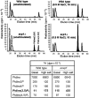
svp1Δ cells accumulate abnormally large amounts of PtdIns(3,5)P2. Anion-exchange HPLC chromatograms of the PtdInsP and PtdInsP2 complements of wild-type and svp1Δ cells, and their responses to hyperosmotic stress. The deacylated phosphoinositides eluted in the order: PtdIns3P, PtdIns4P, PtdIns(3,5)P2 (filled peak) and PtdIns(4,5)P2 (see lower left panel). The relative amounts of phosphoinositides in svp1Δ cells are also shown in the table. The total phosphoinositide complement is unchanged, but in svp1Δ cells an abnormally large proportion of this is PtdIns(3,5)P2. Cells were labelled to isotopic equilibrium, so relative phosphoinositide concentrations match the relative levels of labelling. The data are representative of at least four experiments.
Figure 7
Figure 7
The role of Svp1p in autophagy is PtdIns(3,5)P2-independent, and S. cerevisiae Svp1p-like proteins are not needed for several FAB1/PtdIns(3,5)P2-dependent processes. (A) Autophagy processed Pho8Δ60p normally in fab1Δ cells, but not in svp1Δ cells or in the autophagy mutant apg1Δ (for details, see Materials and methods). (B) Svp1p-related proteins are not needed to maintain growth at elevated temperatures (for details, see Materials and methods). All strains grew at 23°C, so only the 42°C plate is shown. Data are representative of those from three or four experiments that gave similar results. (C) Svp1p-related proteins are not needed for vacuole acidification, as assessed by accumulation of the fluorescent weak base quinacrine. (D) Svp1p-related proteins are not needed for the sorting of proteins into MVB, as assessed by the trafficking of GFP-Phm5p.
Figure 8
Figure 8
The involvement of Fab1p, PtdIns(3,5)P2 and downstream effector proteins in yeast cell functions. (A) Outline of the cycle of vacuole membrane addition and retrieval for which PtdIns(3,5)P2 appears to be essential. It is not clear whether retrograde vacuole-to-late-endosome trafficking occurs by re-segregation of the vacuole and late endosome or by the traffic of a vesicular intermediate between these structures. (B) A tentative synthesis of how the actions of multiple PtdIns(3,5)P2 effector proteins may contribute to various cell functions (see Discussion). All of these, except the involvement of Svp1p in autophagy, require the presence in cells of FAB1 and/or PtdIns(3,5)P2.

Similar articles

Cited by

References

    1. Abeliovich H, Zhang C, Dunn WA Jr, Shokat KM, Klionsky DJ (2003) Chemical genetic analysis of Apg1 reveals a non-kinase role in the induction of autophagy. Mol Biol Cell 14: 477–490 - PMC - PubMed
    1. Baginski ES, Foa PP, Zak B (1967) Microdetermination of inorganic phosphate, phospholipids, and total phosphate in biologic materials. Clin Chem 13: 326–332 - PubMed
    1. Barth H, Meiling-Wesse K, Epple UD, Thumm M (2001) Autophagy and the cytoplasm to vacuole targeting pathway both require Aut10p. FEBS Lett 508: 23–28 - PubMed
    1. Barth H, Meiling-Wesse K, Epple UD, Thumm M (2002) Mai1p is essential for maturation of proaminopeptidase I but not for autophagy. FEBS Lett 512: 173–179 - PubMed
    1. Bonangelino CJ, Catlett NL, Weisman LS (1997) Vac7p, a novel vacuolar protein, is required for normal vacuole inheritance and morphology. Mol Cell Biol 17: 6847–6858 - PMC - PubMed

Publication types

MeSH terms

Substances