. 2004 May 19;23(10):2083-91.
doi: 10.1038/sj.emboj.7600209. Epub 2004 Apr 22.

In vivo activation of PPAR target genes by RXR homodimers

Affiliations

In vivo activation of PPAR target genes by RXR homodimers

Annemieke IJpenberg et al. EMBO J. .

Abstract

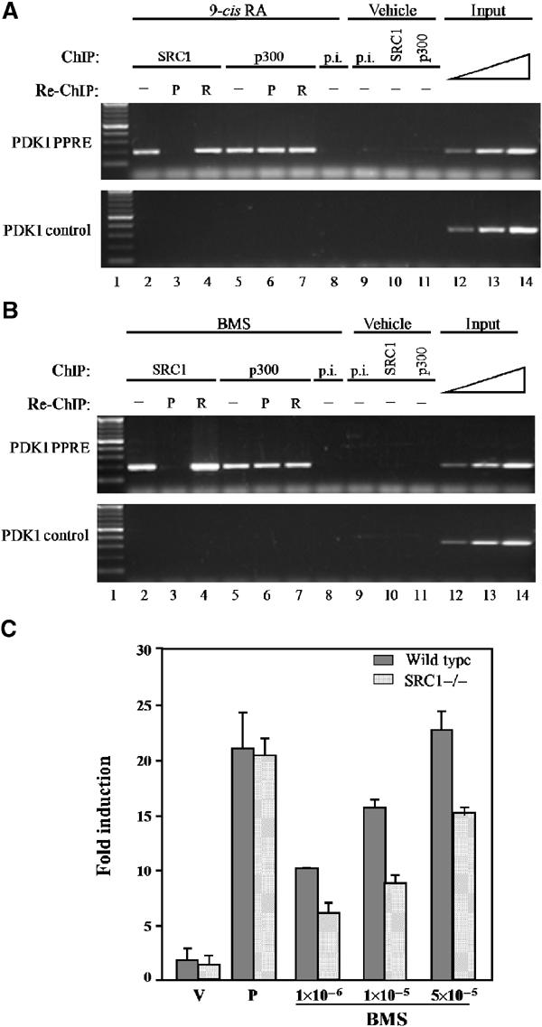
The ability of a retinoid X receptor (RXR) to heterodimerize with many nuclear receptors, including LXR, PPAR, NGF1B and RAR, underscores its pivotal role within the nuclear receptor superfamily. Among these heterodimers, PPAR:RXR is considered an important signalling mediator of both PPAR ligands, such as fatty acids, and 9-cis retinoic acid (9-cis RA), an RXR ligand. In contrast, the existence of an RXR/9-cis RA signalling pathway independent of PPAR or any other dimerization partner remains disputed. Using in vivo chromatin immunoprecipitation, we now show that RXR homodimers can selectively bind to functional PPREs and induce transactivation. At the molecular level, this pathway requires stabilization of the homodimer-DNA complexes through ligand-dependent interaction with the coactivator SRC1 or TIF2. This pathway operates both in the absence and in the presence of PPAR, as assessed in cells carrying inactivating mutations in PPAR genes and in wild-type cells. In addition, this signalling pathway via PPREs is fully functional and can rescue the severe hypothermia phenotype observed in fasted PPARalpha-/- mice. These observations have important pharmacological implications for the development of new rexinoid-based treatments.


Figures

Figure 1
Figure 1
Specific interaction of SRC1 with DNA-bound RXR:RXR complexes. (A) EMSA: Radiolabelled MEp was incubated with RXRα, PPARγ and SRC1(B), in the presence or absence of 1 μM 9-cis RA or 5 μM BRL49653, as indicated. The arrowheads indicate bound PPAR:RXR (grey), RXR:RXR (open) and ternary SRC1(B)-containing complex (black). Ab: polyclonal PPAR antibody; p.i.: preimmune serum. (B) Pull-down assays: Different combinations of RXRα, PPARγ, and either full-length SRC1 or the SRC1 nuclear receptor interacting domain (SRC1(B)) were incubated in 96-well plates coated with biotinylated DNA, either MEp-biotin or PDK1-biotin, in the presence of 40 μM 9-cis RA and/or 10 μM Rosiglitazone. After washing of the wells, retained proteins were analysed by Western blot with antibodies directed against GST (recognizing the GST-SRC1(B) or SRC1 (recognizing the full-length SRC1) as indicated. (C) EMSA: Analyses were performed as in (A), but in the absence of PPAR. DR1G: synthetic RXRE; MEp: functional PPRE from the malic enzyme promoter; AcoA: functional PPRE from the acyl-CoA oxidase promoter; HD: functional PPRE from the bifunctional enzyme promoter; MEd: nonfunctional DR1 element in the malic enzyme promoter.
Figure 2
Figure 2
RXR antagonists preclude RXR:RXR/SRC-1 complex formation. (A) EMSA: Radiolabelled MEp (left panel) or the synthetic RXRE DR1G (right panel) was incubated with RXRα and SRC1(B) in the presence or absence of 1 μM 9-cis RA, LG100268 or LG100754 as indicated. The open arrowhead indicates the RXR homodimer complex, and the black arrowhead indicates the ternary complex. (B) NIH-3T3 cells were transfected with the MEp-containing reporter gene, RXR encoding expression vector, and pCMV-β-galactosidase as internal control for transfection efficiency. Following transfection, the cells were cultivated for 48 h in the presence of 1 μM 9-cis RA, LG100268 or LG100754 as indicated, or with vehicle alone. CAT activity levels were standardized for β-galactosidase expression and expressed as fold induction with respect to the CAT activity obtained in the presence of the vehicle alone. (C) NIH-3T3 cells were transfected with the MEp-containing reporter gene and either an RXR- or a PPAR-encoding expression vector as indicated, together with pCMV-β-galactosidase as internal control for transfection efficiency. A vector expressing a dominant-negative form of SRC1 (SRCdn) was added at increasing doses, as indicated; total transfected DNA was maintained equal under each condition by use of the corresponding empty vector. Numbers indicate the relative proportions (μg) of transfected expression vectors. Following transfection, the cells were cultivated for 48 h in the absence or presence of either 1 μM 9-cis RA, 100 μM Wy14,643, 5 μM BRL49653 or vehicle alone. CAT activity levels were standardized for β-galactosidase expression and expressed as fold induction with respect to the activity observed in the presence of vehicle alone.
Figure 3
Figure 3
Specificity of RXR/coactivator association. EMSA: Radiolabelled MEp was incubated with PPARγ and RXRα (A), or RXR alone (B) and the receptor interacting domains of either SRC1 (amino acids 1226–1441; S), TIF2 (amino acids 624–869; T) or p300 (amino acids 31–220; 300), in the presence or absence of 1 μM 9-cis RA or 5 μM BRL49643 as indicated. The open arrowhead indicates the RXR homodimer, and the black arrowhead indicates the ternary complex.
Figure 4
Figure 4
In vivo cofactor-specific binding of RXR homodimers to PPREs. Experiments were performed using primary keratinocyte cultures derived from either wild-type mice (WT), compound knock-out mice for PPARα and PPARβ (PPARα−/−β−/−), and mice carrying a keratinocyte-specific deletion of RXRα (RXRα−/−). Primary cultures were transfected with a PPRE-luciferase reporter gene and treated with either PPAR ligands (P; a mixture of 10 μM Wy14,643 and 5 μM L1165041), RXR ligand (X; 0.5 μM 9-cis RA) or vehicle (V; DMSO), and subjected to analyses. (A) Schematic outline of the procedure. (B) Reporter gene induction in response to various ligands. Luciferase activity levels were standardized for β-galactosidase expression used as internal control of transfection efficiency, and expressed as the fold induction with respect to the activity observed with the control reporter gene. (C) PPAR and RXR binding to PPREs in vivo. Chromatin from the various primary keratinocyte cultures was immunoprecipitated with either a pan-PPAR antibody (PPAR-Ab), an RXR antibody (RXR-Ab) or preimmune serum (p.i.). Enrichment of either a DNA fragment encompassing the PPRE from the transfected reporter gene (rep PPRE), a PPRE-containing fragment from the endogenous PDK1 promoter (PDK1 PPRE) or a control DNA fragment from the PDK1 gene (PDK1 control) was evaluated by PCR. Aliquots of the extracts were also analysed before immunoprecipitation (input). (D) Ligand-dependent coactivator association in solution. Western blot analyses were performed on proteins after immunoprecipitation (IP) with either an antibody against PPAR (PPAR-Ab) or RXR (RXR-Ab). Proteins were separated by SDS–PAGE, blotted and probed with either SRC1 or TIF2 primary antibody as indicated. Bands were detected by chemiluminescence with horseradish peroxidase. The Western blot control with tubulin antibody was performed on cell lysates prior to immunoprecipitation. (E) SRC1 binds to DNA-bound RXR, in a 9-cis RA-dependent manner. ChIP of PPRE-containing genes was performed as described in (C), using anti-SRC1 or preimmune serum (p.i.).
Figure 5
Figure 5
RXR:RXR/SRC1 ternary complexes bind to PPREs in the presence of PPAR. Primary keratinocytes from wild-type mice were treated with either 0.5 μM 9-cis RA (A), 1 μM BMS 649 (B) or vehicle. Following immunoprecipitation (ChIP) with either an SRC1-specific antibody (SRC1), a p300-specific antibody (p300) or preimmune serum (p.i.), the complexes were subjected to a second immunoprecipitation (re-ChIP) with either a PPAR- (P) or an RXR- (R) specific antibody. Enrichment of a PPRE-containing fragment from the endogenous PDK1 promoter (PDK1 PPRE) and a control DNA fragment from the PDK1 gene (PDK1 control) was evaluated by PCR. Aliquots of the extracts were also analysed before immunoprecipitation (input). (C) Primary keratinocytes from either wild-type (grey bars) or SRC1−/− (hatched bars) mice were transfected with a PPRE-luciferase reporter gene and treated with either vehicle (V; DMSO), PPAR ligands (P; a mixture of 10 μM Wy14,643 and 5 μM L1165041) or increasing doses of BMS as indicated. Luciferase activity levels were standardized for β-galactosidase expression used as internal control of transfection efficiency and expressed as the fold induction with respect to the activity observed with the activity of the reporter gene in the presence of vehicle alone.
Figure 6
Figure 6
RXR homodimers are functional in vivo. (A) SRC1 and TIF2 expression in the liver. Western blot analysis of SRC-1 and TIF2 protein expression levels in a liver protein extract. For the purpose of comparison and control, a protein extract from pup skin was used. Equal loading was verified by β-tubulin levels. (B) PPAR and RXR interaction with TIF2 in solution. Wild-type (WT) and PPARα−/− (KO) mice were fed for 5 days with either Wy14,643 (50 mg/kg/day), 9-cis RA (30 mg/kg/day) or vehicle (solvent). Western blot analyses were performed on protein extracts from the liver and subjected to immunoprecipitation (IP) with either an antibody against PPAR (PPAR-Ab) or RXR (RXR-Ab). Proteins were separated by SDS–PAGE, blotted and probed with a TIF2 primary antibody. Bands were detected by chemiluminescence with horseradish peroxidase. The Western blot control with tubulin antibody was performed on cell lysates prior to immunoprecipitation. (C) ChIP of MEp with PPAR, RXR and TIF2 antibodies. Wild-type (WT) and PPARα−/− (KO) mice were fed for 5 days with either Wy14,643 (50 mg/kg/day), 9-cis RA (30 mg/kg/day) or vehicle (solvent). ChIP of liver extracts was performed with either a PPARα (PPAR-Ab), RXRα (RXR-Ab) or TIF2 (TIF2-Ab) antibody as indicated, and analysed by PCR for enrichment of the MEp element (top panels) or a control sequence (bottom panels). p.i.: preimmune serum. (D) Evaluation of ME gene activity in livers of mice treated with various ligands. Wild-type (WT) or PPARα KO (KO) mice were treated for 5 days with either a PPARα ligand (Wy: Wy14,643, 50 mg/kg/day), RXR ligand (9-cis RA, 30 mg/kg/day), PPARβ ligand (LD: L165041, 30 mg/kg/day), PPARγ ligand (Tro: troglitazone, 250 mg/kg/day), RAR ligand TTNPB (3 μg/kg/day) or vehicle (solvent, 0.5% carboxymethylcellulose), as indicated. Total liver RNA was isolated and expression levels of the ME and L27 genes were analysed by RNase protection assay using gene-specific probes. The indicated ratio of ME to L27 expression levels was determined for each of the samples. An arbitrary value of 1 was assigned to the value ratio obtained in mice treated with the solvent alone. (E) ChIP of the apoCIII DR1 element, in the liver. Wild-type (WT) and PPARα−/− (KO) mice were fed for 5 days with either Wy14,643 (50 mg/kg/day), 9-cis RA (30 mg/kg/day) or vehicle (solvent). ChIP of liver extracts was performed with either a PPARα (PPAR-Ab), RXRα (RXR-Ab) or TIF2 (TIF2-Ab) antibody as indicated, and analysed by PCR for enrichment of the apoCIII DR1 element (top panels) or a control sequence (bottom panels). p.i.: preimmune serum.
Figure 7
Figure 7
Activation of the RXR homodimer signalling pathway prevents fasting-induced hypothermia. Following a 5-day treatment with 9-cis RA (30 mg/kg/day) or vehicle (V), wild-type (circle) and PPARα−/− (triangle) mice were subjected to an overnight fast. Each circle corresponds to the rectal temperature of an individual mouse. Rectal temperatures under fasting conditions are shown on the left, and rectal temperatures of similar groups of mice measured under nonfasting conditions are shown on the right.

Similar articles

Cited by

References

    1. Berger J, Leibowitz MD, Doebber TW, Elbrecht A, Zhang B, Zhou G, Biswas C, Cullinan CA, Hayes NS, Li Y, Tanen M, Ventre J, Wu MS, Berger GD, Mosley R, Marquis R, Santini C, Sahoo SP, Tolman RL, Smith RG, Moller DE (1999) Novel peroxisome proliferator-activated receptor (PPAR) gamma and PPARdelta ligands produce distinct biological effects. J Biol Chem 274: 6718–6725 - PubMed
    1. Desvergne B, Wahli W (1999) Peroxisome proliferator-activated receptors: nuclear control of metabolism. Endocr Rev 20: 649–688 - PubMed
    1. Di-Poï N, Tan NS, Michalik L, Wahli W, Desvergne B (2002) Antiapoptotic role of PPARbeta in keratinocytes via transcriptional control of the Akt1 signaling pathway. Mol Cell 10: 721–733 - PubMed
    1. Dilworth FJ, Fromental-Ramain C, Remboutsika E, Benecke A, Chambon P (1999) Ligand-dependent activation of transcription in vitro by retinoic acid receptor alpha/retinoid X receptor alpha heterodimers that mimics transactivation by retinoids in vivo. Proc Natl Acad Sci USA 96: 1995–2000 - PMC - PubMed
    1. DiRenzo J, Soderstrom M, Kurokawa R, Ogliastro MH, Ricote M, Ingrey S, Horlein A, Rosenfeld MG, Glass CK (1997) Peroxisome proliferator-activated receptors and retinoic acid receptors differentially control the interactions of retinoid X receptor heterodimers with ligands, coactivators, and corepressors. Mol Cell Biol 17: 2166–2176 - PMC - PubMed

Publication types