. 2004 May 19;23(10):2105-15.
doi: 10.1038/sj.emboj.7600216. Epub 2004 Apr 22.

The hydrogenase-like Nar1p is essential for maturation of cytosolic and nuclear iron-sulphur proteins

Affiliations

The hydrogenase-like Nar1p is essential for maturation of cytosolic and nuclear iron-sulphur proteins

Janneke Balk et al. EMBO J. .

Abstract

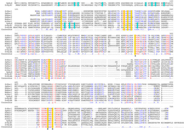
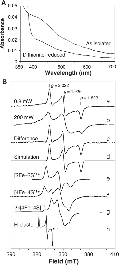
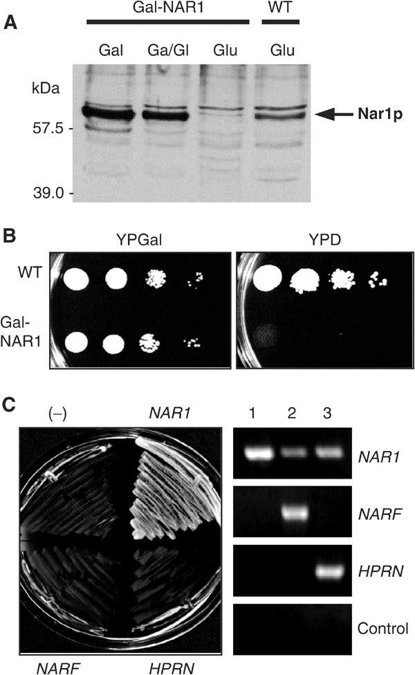
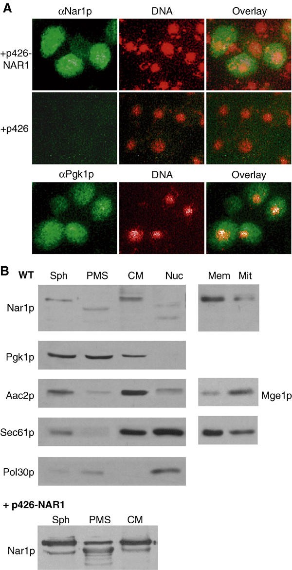
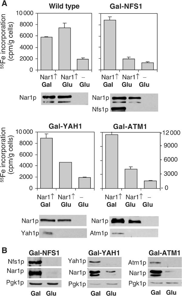
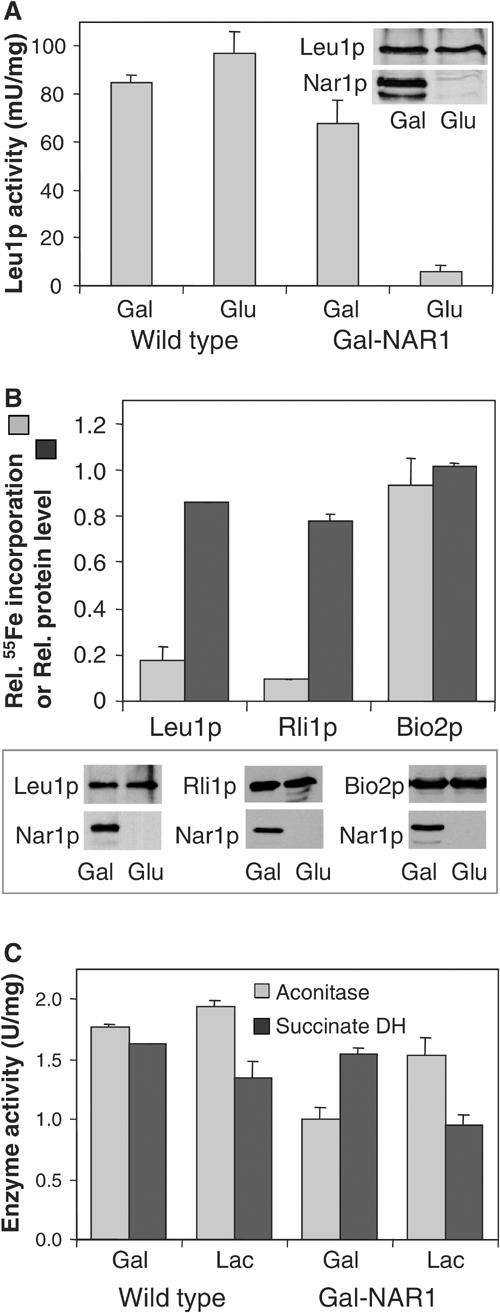
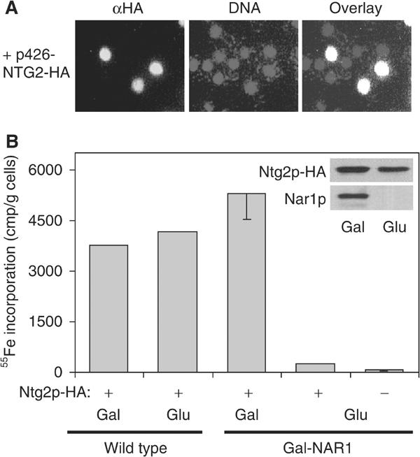
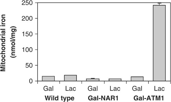
The genome of the yeast Saccharomyces cerevisiae encodes the essential protein Nar1p that is conserved in virtually all eukaryotes and exhibits striking sequence similarity to bacterial iron-only hydrogenases. A human homologue of Nar1p was shown previously to bind prenylated prelamin A in the nucleus. However, yeast neither exhibits hydrogenase activity nor contains nuclear lamins. Here, we demonstrate that Nar1p is predominantly located in the cytosol and contains two adjacent iron-sulphur (Fe/S) clusters. Assembly of its Fe/S clusters crucially depends on components of the mitochondrial Fe/S cluster biosynthesis apparatus such as the cysteine desulphurase Nfs1p, the ferredoxin Yah1p and the ABC transporter Atm1p. Using functional studies in vivo, we show that Nar1p is required for maturation of cytosolic and nuclear, but not of mitochondrial, Fe/S proteins. Nar1p-depleted cells do not accumulate iron in mitochondria, distinguishing these cells from mutants in components of the mitochondrial Fe/S cluster biosynthesis apparatus. In conclusion, Nar1p represents a crucial, novel component of the emerging cytosolic Fe/S protein assembly machinery that catalyses an essential and ancient process in eukaryotes.


Figures

Figure 1
Figure 1
Sequence alignment of Nar1p-like proteins and iron-only hydrogenases. The multisequence alignment of Nar1p-like proteins, Desulfovibrio HydA and HydC, and clostridial CpI hydrogenases was performed using the Multalin program (Corpet, 1988). Conserved cysteine residues are highlighted in yellow and cyan (hydrogenase-specific). The cysteine residues coordinating the H-cluster in iron-only hydrogenases are indicated (H). Abbreviations for organisms: Sc, S. cerevisiae; Sp, Schizosaccharomyces pombe; Hs, Homo sapiens; Mm, Mus musculus; At, Arabidopsis thaliana; Cp; Clostridium pasteurianum; Dv; Desulfovibrio vulgaris.
Figure 2
Figure 2
Spectroscopic analysis of purified Nar1p. (A) Absorption spectra of Strep-tagged Nar1p (1.6 μM) as isolated and after reduction with 1 mM sodium dithionite in buffer TNE (100 mM Tris–HCl, pH 8, 150 mM NaCl, 1 mM EDTA) containing 40 μM desthiobiotin. (B) EPR spectra of recombinant Nar1p were recorded under the following conditions: microwave frequency, 9.460±0.001 GHz; modulation frequency, 100 kHz; modulation amplitude, 1.25 mT. (a) Nar1p (50 μM) reduced with 5 mM sodium dithionite in buffer TNE with 1 mM desthiobiotin (0.80 mW, 10 K). (b) As in (a), but recorded at 200 mW, 10 K. (c) The sharp rhombic EPR signal obtained by subtracting an appropriate intensity of spectrum b from a. (d) A theoretical simulation of spectrum c (Beinert and Albracht, 1982). Simulation parameters, gz=2.022, gy=1.926 and gx=1.823; linewidths 2.5, 3.0 and 3.1 mT, respectively. For comparison, traces (e–h) were added with appropriate corrections of the magnetic fields to the microwave frequency of Nar1p experiments. (e) [2Fe–2S]1+ cluster in reduced spinach leaf ferredoxin (Hagen and Albracht, 1982). (f) [4Fe–4S]1+ cluster in partially dithionite-reduced heterodisulphide reductase from Methanothermobacter marburgensis (9.459 GHz, 20 mW, 30 K; courtesy of Dr R Hedderich (MPI for Terrestrial Microbiology, Marburg). (g) Two interacting [4Fe–4S]1+ clusters in fully reduced ferredoxin from Acidaminococcus fermentans (9.65 GHz, 2 mW, 12 K; Thamer et al, 2003). (h) Oxidised H-cluster in thionine-treated C. pasteurianum Fe-hydrogenase CpI (9.23 GHz, 10 mW, 20 K; Adams, 1987).
Figure 3
Figure 3
Yeast NAR1 is an essential gene and cannot be replaced by its human homologues. (A) Wild-type (WT) and Gal-NAR1 cells were grown in liquid rich medium supplemented with galactose (Gal), a 1:1 mixture of galactose and glucose (Ga/Gl) or glucose (Glu). A total cell extract prepared by alkaline lysis was subjected to immunostaining for Nar1p using an anti-Nar1p antiserum. The minor band at 57 kDa may represent a breakdown product of mature Nar1p. (B) Wild-type and Gal-NAR1 cells were grown for 2 × 2 days at 30°C on agar plates containing rich media supplemented with galactose (YPGal) or glucose (YPD). 10-fold serial dilutions are shown. (C) Gal-NAR1 cells were transformed with plasmid p416MET25 containing either no insert (−), the yeast NAR1 gene, or the genes of human NARF and HPRN. Cells were grown on YPD medium for 2 × 2 days at 30°C (left). Expression of the human genes was tested by RT–PCR using galactose-grown cells harbouring plasmids with (1) yeast NAR1, (2) NARF and (3) HPRN genes. A control reaction was performed without reverse transcriptase.
Figure 4
Figure 4
Subcellular localisation of Nar1p. (A) In situ localisation of Nar1p. Wild-type cells (strain PSY581) were transformed with the high-copy expression vector p426 containing NAR1 or no gene. Log-phase cells were fixed with 2.4% (w/v) formaldehyde, permeabilised and labelled with purified anti-Nar1p (αNar1p) or monoclonal anti-Pgk1p (αPgk1p) antibodies, followed by fluorophore-conjugated secondary antibodies (green). DNA was counterstained with DAPI and is indicated in red. Similar results with low fluorescence intensity were obtained with NAR1 inserted into low-copy expression vector p416MET25. (B) Immunoblot analysis of Nar1p in cell fractions. Log-phase wild-type cells were converted into spheroplasts (Sph), subjected to Dounce homogenisation and, after removal of intact cells and cell debris, the extract was fractionated into post-mitochondrial supernatant (PMS) and crude mitochondria (CM) by centrifugation for 10 min at 12 000 g. A similar fractionation was performed with cells overproducing Nar1p from vector p426-NAR1. A separate wild-type yeast culture was used to isolate nuclei (Nuc) by Ficoll gradient centrifugation (Aris and Blobel, 1991). Equal amounts of protein (20 μg/per lane) were separated by SDS–PAGE, blotted and immunostained for Nar1p or marker proteins of known cellular localisation (left panel; cytosolic phosphoglycerate kinase Pgk1p; mitochondrial ATP/ADP carrier Aac2p; endoplasmic reticulum translocon subunit Sec61p; and nuclear DNA polymerase-associated Pol30p). The crude mitochondrial fraction was further separated on a Nycodenz step-gradient into fractions containing enriched mitochondria (Mit) and microsomal membranes (Mem). Samples were analysed by immunostaining (right panel; Mge1p; mitochondrial matrix).
Figure 5
Figure 5
Fe/S cluster assembly on Nar1p depends on the mitochondrial ISC machinery. (A) Wild-type and the galactose-regulatable Gal-NFS1, Gal-YAH1 and Gal-ATM1 cells were transformed with plasmid p426-NAR1 (Nar1↑) or empty vector p426 (−). Cells were grown in iron-poor medium supplemented with galactose (Gal) or glucose (Glu) for 24 h. Cells were radiolabelled with 55Fe and disrupted with glass beads in Triton X-100-containing buffer. Nar1p was isolated from the cell extract by immunoprecipitation and the amount of 55Fe associated with Nar1p was quantified by scintillation counting. The indicated proteins were visualised by immunostaining of cell extracts. (B) The indicated cells overproducing Nar1p were grown for 40 h and cell extracts were analysed as in (A).
Figure 6
Figure 6
Nar1p is required for maturation of cytosolic, but not of mitochondrial, Fe/S proteins. (A) Wild-type and Gal-NAR1 cells were grown in minimal medium supplemented with galactose (Gal) or glucose (Glu). The enzyme activity of the cytosolic isopropylmalate isomerase (Leu1p) was estimated in total cell extracts. The levels of Leu1p and Nar1p in Gal-NAR1 cells were measured by immunoblot analysis (inset). (B) Gal-NAR1 cells were grown in iron-poor medium supplemented with galactose or glucose. Cells were transformed with high-copy plasmids carrying the genes for an HA-tagged version of cytosolic Rli1p or for mitochondrial Bio2p. Radiolabelling with 55Fe, preparation of cell extracts and immunoprecipitation of the Fe/S protein of interest were performed as in Figure 5. For Leu1p, the endogenous protein was analysed. Results are given as the ratio of 55Fe incorporation in Nar1p-depleted (glucose-grown) cells and that in Nar1p-expressing (galactose-grown) cells, corrected for a small background (<2% of total signal of galactose-grown cells). Protein levels of the indicated proteins were determined by immunoblot analysis (lower panel) and quantified by densitometry (upper panel). (C) The enzyme activities (in U per mg of mitochondrial protein) of aconitase and succinate dehydrogenase (DH) were assayed in mitochondria isolated from wild-type or Gal-NAR1 cells after growth in minimal medium containing galactose or lactate (Lac).
Figure 7
Figure 7
Maturation of the nuclear Fe/S protein Ntg2p depends on Nar1p function. Wild-type or Gal-NAR1 cells were transformed with a high-copy plasmid (p426-NTG2-HA) encoding an HA-tagged version of Ntg2p. (A) Localisation of overproduced HA-tagged Ntg2p by immunofluorescence was performed as in Figure 4A. (B) After growth of cells in galactose- (Gal) or glucose-containing (Glu) iron-poor minimal medium, maturation of Ntg2p was measured by following the 55Fe incorporation by immunoprecipitation with anti-HA antibodies as described in Figure 5A. A control immunoprecipitation was performed with cells lacking HA-tagged Ntg2p (−). The inset shows immunostaining of Ntg2p-HA and Nar1p in extracts of Gal-NAR1 cells.
Figure 8
Figure 8
Depletion of Nar1p does not lead to iron accumulation in mitochondria. Wild-type, Gal-NAR1 and Gal-ATM1 cells were grown for 40 h in minimal medium containing galactose (Gal) or in lactate medium (Lac). Mitochondria were isolated and the amount of total iron was determined.

Similar articles

Cited by

References

    1. Adams MW (1987) The mechanisms of H2 activation and CO binding by hydrogenase I and hydrogenase II of Clostridium pasteurianum. J Biol Chem 262: 15054–15061 - PubMed
    1. Alseth I, Eide L, Pirovano M, Rognes T, Seeberg E, Bjoras M (1999) The Saccharomyces cerevisiae homologues of endonuclease III from Escherichia coli, Ntg1 and Ntg2, are both required for efficient repair of spontaneous and induced oxidative DNA damage in yeast. Mol Cell Biol 19: 3779–3787 - PMC - PubMed
    1. Aris JP, Blobel G (1991) Isolation of yeast nuclei. Methods Enzymol 194: 735–749 - PubMed
    1. Barton RM, Worman HJ (1999) Prenylated prelamin A interacts with Narf, a novel nuclear protein. J Biol Chem 274: 30008–30018 - PubMed
    1. Beinert H, Albracht SPJ (1982) New insights, ideas and unanswered questions concerning iron–sulfur clusters in mitochondria. Biochim Biophys Acta 683: 245–277 - PubMed

Publication types

MeSH terms