. 2004 May 19;23(10):2116-25.
doi: 10.1038/sj.emboj.7600217. Epub 2004 Apr 22.

Phosphorylation-dependent degradation of c-Myc is mediated by the F-box protein Fbw7

Affiliations

Phosphorylation-dependent degradation of c-Myc is mediated by the F-box protein Fbw7

Masayoshi Yada et al. EMBO J. .

Abstract

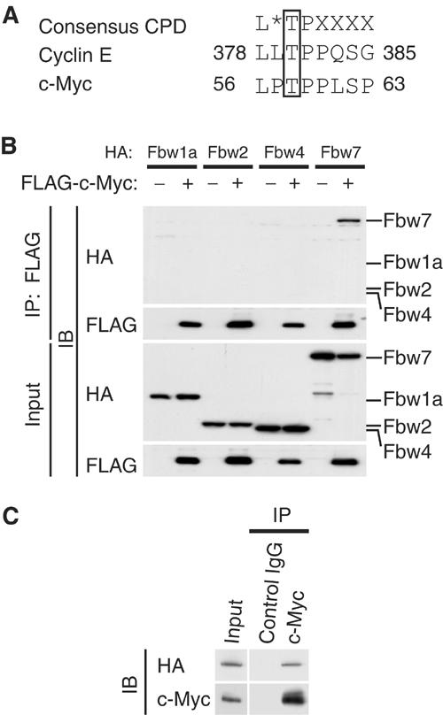
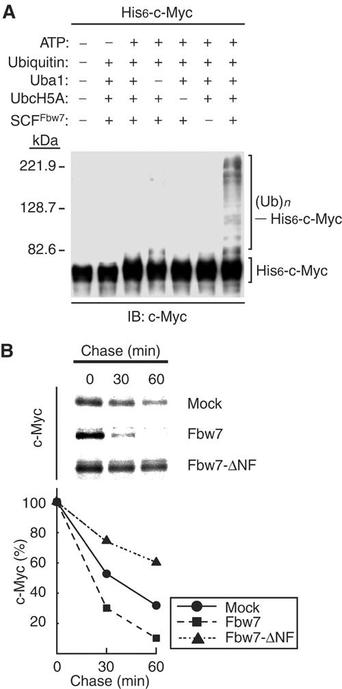
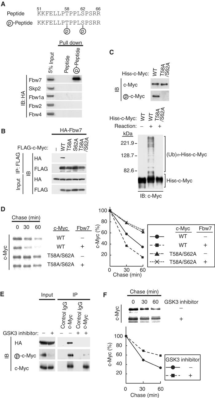
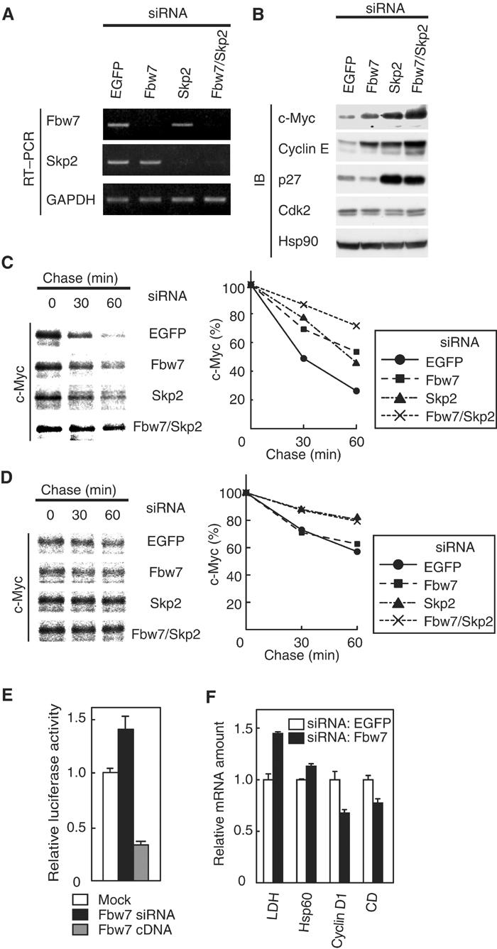
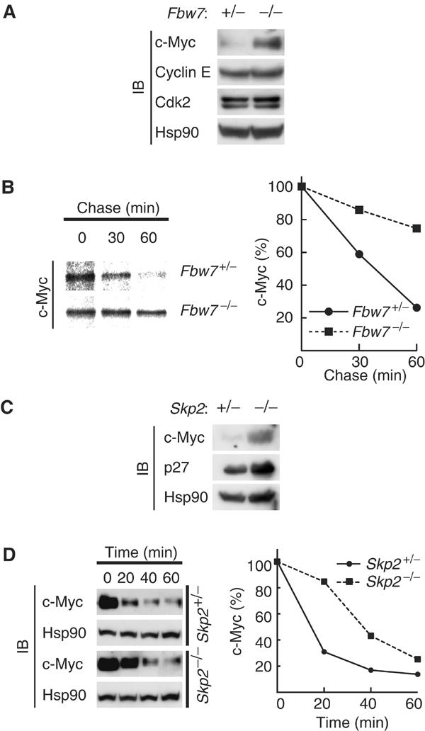
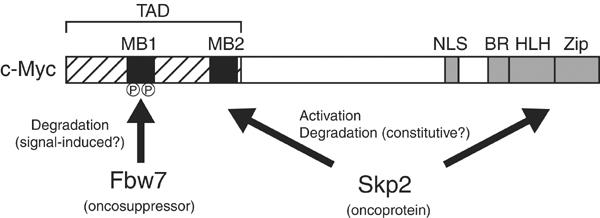
The F-box protein Skp2 mediates c-Myc ubiquitylation by binding to the MB2 domain. However, the turnover of c-Myc is largely dependent on phosphorylation of threonine-58 and serine-62 in MB1, residues that are often mutated in cancer. We now show that the F-box protein Fbw7 interacts with and thereby destabilizes c-Myc in a manner dependent on phosphorylation of MB1. Whereas wild-type Fbw7 promoted c-Myc turnover in cells, an Fbw7 mutant lacking the F-box domain delayed it. Furthermore, depletion of Fbw7 by RNA interference increased both the abundance and transactivation activity of c-Myc. Accumulation of c-Myc was also apparent in mouse Fbw7-/- embryonic stem cells. These observations suggest that two F-box proteins, Fbw7 and Skp2, differentially regulate c-Myc stability by targeting MB1 and MB2, respectively.


Figures

Figure 1
Figure 1
Interaction of Fbw7 with c-Myc in vivo. (A) The Cdc4 phospho-degron (CPD) sequences of human cyclin E and c-Myc. The asterisk indicates L, I, or P; X indicates any residue other than K or R. (B) HEK293T cells were transfected with vectors for FLAG-c-Myc and either HA-Fbw1a, HA-Fbw2, HA-Fbw4, or HA-Fbw7, as indicated, and were then incubated with MG132 for 6 h. Cell lysates were subjected to immunoprecipitation (IP) with antibodies to FLAG, and the resulting precipitates as well as the original cell lysates (input) were subjected to immunoblot analysis (IB) with antibodies to HA or FLAG. (C) HEK293T cells were transfected with a vector for HA-Fbw7, subjected to serum deprivation, stimulated by re-exposure to serum for 2 h, and then incubated for 6 h in the additional presence of MG132. Cell lysates were then subjected to immunoprecipitation with antibodies to c-Myc or with control mouse IgG, and the resulting precipitates were subjected to immunoblot analysis with antibodies to HA or c-Myc.
Figure 2
Figure 2
Promotion of the ubiquitylation and degradation of c-Myc by Fbw7. (A) Ubiquitylation of c-Myc by the recombinant SCFFbw7 complex in vitro. Recombinant SCFFbw7 was assayed for ubiquitylation activity with His6-c-Myc as substrate in the absence or presence of the indicated reaction mixture components. The reaction mixtures were then subjected to immunoblot analysis with antibodies to c-Myc. The positions of unmodified His6-c-Myc and of His6-c-Myc conjugated with ubiquitin ((Ub)n) are indicated. (B) Promotion of c-Myc degradation by Fbw7 in vivo. HEK293T cells were transfected with vectors for FLAG-c-Myc and either HA-Fbw7 or HA-Fbw7-ΔNF (or the corresponding empty vector; mock). The cells were then subjected to pulse-chase analysis by metabolic labeling with [35S]methionine, and cell lysates were prepared at the indicated times of the chase incubation and subjected to immunoprecipitation with antibodies to FLAG. The precipitates were subjected to SDS–polyacrylamide gel electrophoresis and autoradiography (upper panel). The percentage of FLAG-c-Myc remaining after the various chase times was quantitated by image analysis (lower panel).
Figure 3
Figure 3
Phosphorylation of c-Myc on Thr-58 and Ser-62 is required for its recognition by the SCFFbw7complex. (A) Interaction of Fbw7 with a synthetic CPD peptide in vitro. HEK293T cells were transfected with vectors for HA-Fbw7, HA-Skp2, HA-Fbw1a, HA-Fbw2, or HA-Fbw4. Cell lysates were subsequently subjected to a ‘pull-down' assay with beads linked to nonphosphorylated or phosphorylated peptides corresponding to the CPD of c-Myc (upper panel), and the resulting precipitates (or 5% of the input cell lysates) were subjected to immunoblot analysis with antibodies to HA. (B) In vivo association of Fbw7 with c-Myc derivatives. HEK293T cells were transfected with vectors for HA-Fbw7 and either wild type (WT) or the indicated Thr-58 or Ser-62 mutants of FLAG-c-Myc. They were then subjected to in vivo binding analysis as described in Figure 1B. (C) Ubiquitylation of phosphorylated but not nonphosphorylated c-Myc by recombinant SCFFbw7 in vitro. His6-c-Myc and the His6-c-Myc(T58A/S62A) mutant purified from Sf21 cells were subjected to immunoblot analysis with antibodies to c-Myc or phospho-c-Myc (upper panel). The purified His6-c-Myc and His6-c-Myc(T58A/S62A) proteins were also tested as substrates in the in vitro ubiquitylation assay, performed with all reaction components (lower panel), as described in Figure 2A. (D) HEK293T cells were transfected with the indicated combinations of FLAG-c-Myc and HA-Fbw7 vectors and then subjected to pulse-chase analysis as described in Figure 2B. (E) HEK293T cells transfected with a vector for HA-Fbw7 were deprived of serum and then stimulated with serum in the absence or presence of a GSK3 inhibitor as described in Materials and methods. Cell lysates were then subjected to immunoprecipitation and immunoblot analysis as described in Figure 1B. (F) HEK293T cells transfected with a vector for FLAG-c-Myc were subjected to pulse-chase analysis in the absence or presence of a GSK3 inhibitor.
Figure 4
Figure 4
Depletion of Fbw7 by RNAi induces accumulation of c-Myc and promotes c-Myc-dependent transactivation. (A) At 48 h after transfection of HeLa cells with the indicated siRNAs, the abundance of mRNAs for Fbw7, Skp2, and glyceraldehyde-3-phosphate dehydrogenase (GAPDH; internal standard) was determined by RT–PCR. (B) At 48 h after siRNA transfection, HeLa cell lysates were subjected to immunoblot analysis with antibodies to c-Myc, cyclin E, p27, Cdk2, or Hsp90 (internal standard). (C) At 48 h after siRNA transfection, HeLa cells were transfected with a vector for FLAG-c-Myc and then subjected to pulse-chase analysis. (D) At 48 h after siRNA transfection, HeLa cells were transfected with a vector for FLAG-c-Myc(T58A/S62A) and then subjected to pulse-chase analysis. (E) Activity of a c-Myc-dependent luciferase reporter gene. At 48 h after mock, Fbw7 siRNA, or Fbw7 cDNA transfection, HeLa cells were transfected with a c-Myc-dependent luciferase reporter plasmid (p4 × E-SVP-Luc). The cells were incubated for an additional 24 h and then assayed for relative luciferase activity. Data are means±s.d. of triplicates from a representative experiment. (F) Transcriptional activity of c-Myc target genes. The abundance of transcripts derived from the indicated genes (CD, carboxypeptidase D) was determined by RT and real-time PCR 72 h after siRNA transfection. Data are means±s.d. of triplicates from a representative experiment.
Figure 5
Figure 5
Stabilization of c-Myc in Fbw7−/− ES cells. (A) Lysates of asynchronous Fbw7+/− or Fbw7−/− ES cells were subjected to immunoblot analysis with antibodies to the indicated proteins. (B) ES cells were transfected with a vector for FLAG-c-Myc controlled by the phosphoglycerate kinase gene promoter and then subjected to pulse-chase analysis. (C) Lysates of asynchronous Skp2+/− or Skp2−/− MEFs were subjected to immunoblot analysis with antibodies to c-Myc, p27, or Hsp90. (D) MEFs were deprived of serum, stimulated by re-exposure to serum for 2 h, and then incubated for the indicated times in the additional presence of cycloheximide (50 μg/ml). Cell lysates were subjected to immunoblot analysis with antibodies to c-Myc or Hsp90.
Figure 6
Figure 6
A model for c-Myc ubiquitylation. Two F-box proteins, Fbw7 and Skp2, regulate the turnover of c-Myc. The putative oncosuppressor Fbw7 recognizes c-Myc molecules that are phosphorylated in the MB1 region of the transcriptional activation domain (TAD). The oncoprotein Skp2 recognizes the MB2 and HLH-Zip domains. Skp2 both enhances the transactivation activity of c-Myc and promotes its degradation. NLS, nuclear localization signal; BR, basic region.

Similar articles

Cited by

References

    1. Bahram F, von der Lehr N, Cetinkaya C, Larsson LG (2000) c-Myc hot spot mutations in lymphomas result in inefficient ubiquitination and decreased proteasome-mediated turnover. Blood 95: 2104–2110 - PubMed
    1. Bornstein G, Bloom J, Sitry-Shevah D, Nakayama K, Pagano M, Hershko A (2003) Role of SCFSkp2 ubiquitin ligase in the degradation of p21Cip1 during S-phase. J Biol Chem 278: 25752–25757 - PubMed
    1. Carrano AC, Eytan E, Hershko A, Pagano M (1999) SKP2 is required for ubiquitin-mediated degradation of the CDK inhibitor p27. Nat Cell Biol 1: 193–199 - PubMed
    1. Charrasse S, Carena I, Brondani V, Klempnauer KH, Ferrari S (2000) Degradation of B-Myb by ubiquitin-mediated proteolysis: involvement of the Cdc34–SCF(p45Skp2) pathway. Oncogene 19: 2986–2995 - PubMed
    1. Ciechanover A, DiGiuseppe JA, Schwartz AL, Brodeur GM (1991) Degradation of MYCN oncoprotein by the ubiquitin system. Prog Clin Biol Res 366: 37–43 - PubMed

Publication types

MeSH terms