. 2004:6:35-54.
doi: 10.1251/bpo71. Epub 2004 Mar 19.

Ribosome formation from subunits studied by stopped-flow and Rayleigh light scattering

Affiliations

Ribosome formation from subunits studied by stopped-flow and Rayleigh light scattering

Ayman Antoun et al. Biol Proced Online. 2004.

Abstract

Light scattering and standard stopped-flow techniques were used to monitor rapid association of ribosomal subunits during initiation of eubacterial protein synthesis. The effects of the initiation factors IF1, IF2, IF3 and buffer conditions on subunit association were studied along with the role of GTP in this process. The part of light scattering theory that is essential for kinetic measurements is high-lighted in the main text and a more general treatment of Rayleigh scattering from macromolecules is given in an appendix.


Figures

Fig. 1
Fig. 1. The anti-association activity of IF3.
The extent of 70S initiation complex formation was monitored as a function of time by light scattering after rapid mixing in the stopped-flow instrument of a volume containing 30S subunits, mRNA, GTP and initiation factors as indicated with a volume containing 50S subunits. Time traces obtained with no initiation factors added to the 30S subunits (Panel A), with only IF3 added (Panel B), with IF1 and IF2 added (Panel C) and with IF1, IF2, IF3 and [3H]fMet-tRNAfMet added (Panel D).
Fig. 2
Fig. 2. Rate of initiation in the absence of IF3 monitored by di-peptide formation.
The extent of dipeptide formation was monitored as a function of time after rapid mixing in a quench flow instrument of a volume containing 30S subunits, mRNA, GTP, IF1 and IF2 with an equal volume containing 50S subunits. Initiator tRNA was present either in the 30S or in the 50S mix. Time curves obtained with [3H]fMet-tRNAfMet added with 30S (Panel A), [3H]fMet-tRNAfMet added with 50S (panel B).
Fig. 3
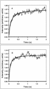
Fig. 3. The effects of G-nucleotides on the association of 30S pre-initiation complex with 50S subunits.
The extent of 70S initiation complex formation was monitored as a function of time by light scattering after rapid mixing of pre-initiation 30S complexes with 50S subunits in a stopped-flow instrument. Traces obtained with GTP (Panel A) and GDP (Panel B).
Fig. 4
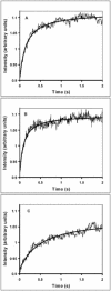
Fig. 4. The effects of buffer composition on the rate of 70S initiation complex formation.
The extent of 70S complex formation was monitored as a function of time by light scattering after rapid mixing of mixture A containing dissociated 70S ribosomes together with IF1, IF3 and mRNA with mixture B containing IF2:GTP together with fMet-tRNA. Complex formation in polymix buffer (PM) with 3 mM of free Mg2+ (Panel A), in PM buffer with 7 mM of free Mg2+ (Panel B) and in PM buffer with 3 mM free Mg2+ plus 10 mM of PEP (Panel C).
Fig. 5
Fig. 5. The effects of the level of magnesium on the rate of naked 70S formation formation from its subunits.
The extent of 70S complex formation was monitored as a function of time by light scattering after rapid mixing of mixture A containing 30S ribosomes with mixture B containing 50S subunits. 70S formation in polymix buffer (PM) with 3 mM of free Mg2+ (Panel A), in PM buffer with 7 mM of free Mg2+ (Panel B).
Fig. 6
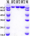
Fig. 6. Purity of His-Tagged IF2 (IF2_HT) as seen on 10% SDS PAGE.
Lanes 1 and 5 contain protein molecular weight marker. Lanes 2, 3, and 4 contain increasing amounts of IF2_HT.

Similar articles

Cited by

References

    1. Gualerzi CO, Pon CL. Initiation of mRNA translation in prokaryotes. Biochemistry. 1990;29:5881–5889. - PubMed
    1. Pestova TV, Hellen CU. The structure and function of initiation factors in eukaryotic protein. Cell Mol Life Sci. 2000;57:651–674. - PMC - PubMed
    1. Roll-Mecak A, Shin BS, Dever TE, Burley SK. Engaging the ribosome: universal IFs of translation. Trends Biochem Sci. 2001;26:705–709. doi: 10.1016/S0968-0004(01)02024-2. - DOI - PubMed
    1. Kyrpides NC, Woese CR. Universally conserved translation initiation factors. Proc Natl Acad Sci USA. 1998;95:224–228. doi: 10.1073/pnas.95.1.224. - DOI - PMC - PubMed
    1. Pestova TV, Kolupaeva VG. The roles of individual eukaryotic translation initiation factors in ribosomal scanning and initiation codon selection. Genes Dev. 2002;16:2906–2922. doi: 10.1101/gad.1020902. - DOI - PMC - PubMed