. 2004 May;48(5):1848-55.
doi: 10.1128/AAC.48.5.1848-1855.2004.

Biochemical characterization of Streptococcus pneumoniae penicillin-binding protein 2b and its implication in beta-lactam resistance

Affiliations

Biochemical characterization of Streptococcus pneumoniae penicillin-binding protein 2b and its implication in beta-lactam resistance

Estelle Pagliero et al. Antimicrob Agents Chemother. 2004 May.

Abstract

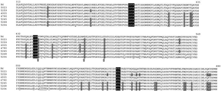
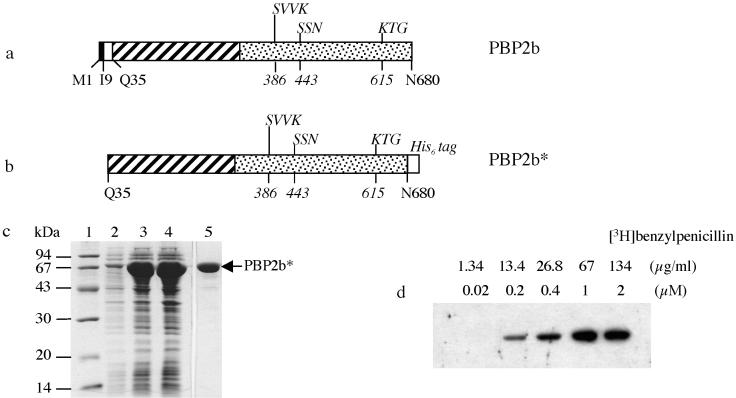
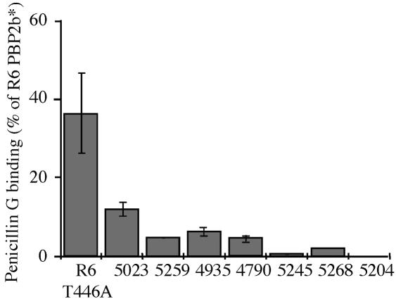
Extensive use of beta-lactam antibiotics has led to the selection of pathogenic streptococci resistant to beta-lactams due to modifications of the penicillin-binding proteins (PBPs). PBP2b from Streptococcus pneumoniae is a monofunctional (class B) high-molecular-weight PBP catalyzing the transpeptidation between adjacent stem peptides of peptidoglycan. The transpeptidase domain of PBP2b isolated from seven clinical resistant (CR) strains contains 7 to 44 amino acid changes over the sequence of PBP2b from the R6 beta-lactam-sensitive strain. We show that the extracellular soluble domains of recombinant PBP2b proteins (PBP2b*) originating from these CR strains have an in vitro affinity for penicillin G that is reduced by up to 99% from that of the R6 strain. The Thr446Ala mutation is always observed in CR strains and is close to the key conserved motif (S(443)SN). The Thr446Ala mutation in R6 PBP2b* displays a 60% reduction in penicillin G affinity in vitro compared to that for the wild-type protein. A recombinant R6 strain expressing the R6 PBP2b Thr446Ala mutation is twofold less sensitive to piperacillin than the parental S. pneumoniae strain. Analysis of the Thr446Ala mutation in the context of the PBP2b CR sequences revealed that its influence depends upon the presence of other unidentified mutations.


Figures

FIG. 1.
FIG. 1.
Alignment of amino acid sequences of the TP domains from residues 313 to 680 of PBP2b from clinical isolates and from the R6 strain. The catalytic motifs of the TP domain are enclosed in dark boxes, and all the mutations present in clinical isolate sequences, compared to the sequence of PBP2b from the R6 strain, are underlined. The mutations present in at least three of the six penicillin-intermediate and penicillin-resistant clinical isolates are underlined and highlighted by shaded boxes. The stars highlight amino acid substitutions present in all clinical isolates: Thr446Ala, Glu476Gly, and Thr489Ser/Ala.
FIG. 2.
FIG. 2.
Expression and purification of PBP2b*R6. (a) Schematic representation of the native PBP2b protein. The solid and empty boxes indicate the cytoplasmic domain and the membrane anchor, respectively. In the periplasmic region, the N-terminal and the TP domains are represented by hatched and dotted boxes, respectively. The conserved motifs of the TP domain are indicated above the boxes (with the first position numbered in italics below). (b) Schematic representation of the construct yielding the soluble form of PBP2b*R6. The His6 tag added in the C terminus of PBP2b* is shown (shadowed box). (c) Expression and purification of PBP2b*R6. Proteins were separated by SDS-12.5% PAGE and stained with Coomassie blue. Lanes: 1, standard molecular mass markers; 2, flowthrough from nickel column; 3, total cell lysate; 4, soluble supernatant following cell lysis; 5, protein eluted at 115 mM imidazole. (d) Saturation binding of PBP2b* by [3H]benzylpenicillin. Fluorogram of a SDS-12.5% PAGE dried gel of PBP2b* (1 μM) labeled with different concentrations of [3H]benzylpenicillin indicated above the gel.
FIG. 3.
FIG. 3.
Relative affinity of PBP2b* proteins for penicillin G. The various purified PBP2b* proteins (0.5 μM) were incubated at 37°C for 30 min with 2 μM [3H]benzylpenicillin. All components were added at the same time, and the reaction was stopped by adding denaturing loading buffer. The affinity for penicillin G for each protein is expressed as a percentage of the PBP2b*R6 value.

References

    1. Chesnel, L., L. Pernot, D. Lemaire, D. Champelovier, J. Croize, O. Dideberg, T. Vernet, and A. Zapun. 2003. The structural modifications induced by the M339F substitution in PBP2x from Streptococcus pneumoniae further decreases the susceptibility to beta-lactams of resistant strains. J. Biol. Chem. 278:44448-44456. - PubMed
    1. Coffey, T. J., M. Daniels, L. K. McDougal, C. G. Dowson, F. C. Tenover, and B. G. Spratt. 1995. Genetic analysis of clinical isolates of Streptococcus pneumoniae with high-level resistance to expanded-spectrum cephalosporins. Antimicrob. Agents Chemother. 39:1306-1313. - PMC - PubMed
    1. Di Guilmi, A. M., A. Dessen, O. Dideberg, and T. Vernet. 2003. The glycosyltransferase domain of penicillin-binding protein 2a from Streptococcus pneumoniae catalyzes the polymerization of murein glycan chains. J. Bacteriol. 185:4418-4423. - PMC - PubMed
    1. Di Guilmi, A. M., A. Dessen, O. Dideberg, and T. Vernet. 2003. Functional characterization of penicillin-binding protein 1b from Streptococcus pneumoniae. J. Bacteriol. 185:1650-1658. - PMC - PubMed
    1. Di Guilmi, A. M., N. Mouz, J. P. Andrieu, J. Hoskins, S. R. Jaskunas, J. Gagnon, O. Dideberg, and T. Vernet. 1998. Identification, purification, and characterization of transpeptidase and glycosyltransferase domains of Streptococcus pneumoniae penicillin-binding protein 1a. J. Bacteriol. 180:5652-5659. - PMC - PubMed

Publication types

MeSH terms