Mutations in a human ROBO gene disrupt hindbrain axon pathway crossing and morphogenesis
- PMID: 15105459
- PMCID: PMC1618874
- DOI: 10.1126/science.1096437
Mutations in a human ROBO gene disrupt hindbrain axon pathway crossing and morphogenesis
Abstract
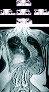
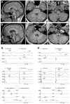
The mechanisms controlling axon guidance are of fundamental importance in understanding brain development. Growing corticospinal and somatosensory axons cross the midline in the medulla to reach their targets and thus form the basis of contralateral motor control and sensory input. The motor and sensory projections appeared uncrossed in patients with horizontal gaze palsy with progressive scoliosis (HGPPS). In patients affected with HGPPS, we identified mutations in the ROBO3 gene, which shares homology with roundabout genes important in axon guidance in developing Drosophila, zebrafish, and mouse. Like its murine homolog Rig1/Robo3, but unlike other Robo proteins, ROBO3 is required for hindbrain axon midline crossing.
Figures


Comment in
-
Neuroscience. Crossing the midline.Science. 2004 Jun 4;304(5676):1455-6. doi: 10.1126/science.1099534. Science. 2004. PMID: 15178787 No abstract available.
References
-
- Seeger M, Tear G, Ferres-Marco D, Goodman CS. Neuron. 1993;10:409. - PubMed
-
- Kidd T, et al. Cell. 1998;92:205. - PubMed
-
- Brose K, et al. Cell. 1999;96:795. - PubMed
-
- Fricke C, Lee JS, Geiger-Rudolph S, Bonhoeffer F, Chien CB. Science. 2001;292:507. - PubMed
-
- Rajagopalan S, Vivancos V, Nicolas E, Dickson BJ. Cell. 2000;103:1033. - PubMed
Publication types
MeSH terms
Substances
Grants and funding
- DC05524/DC/NIDCD NIH HHS/United States
- R01 HL066251/HL/NHLBI NIH HHS/United States
- MH60233/MH/NIMH NIH HHS/United States
- R01 EY015298/EY/NEI NIH HHS/United States
- R01 EY012498/EY/NEI NIH HHS/United States
- EY12498/EY/NEI NIH HHS/United States
- DC00162/DC/NIDCD NIH HHS/United States
- P30 HD018655/HD/NICHD NIH HHS/United States
- EY15298/EY/NEI NIH HHS/United States
- EY13583/EY/NEI NIH HHS/United States
- EY15311/EY/NEI NIH HHS/United States
- R01 EY008313/EY/NEI NIH HHS/United States
- P30 HD 18655/HD/NICHD NIH HHS/United States
- R01 EY013583/EY/NEI NIH HHS/United States
LinkOut - more resources
Full Text Sources
Other Literature Sources
Medical
Molecular Biology Databases

