. 2004 Jun;74(6):1239-48.
doi: 10.1086/421331. Epub 2004 Apr 21.

Mutations in the human TBX4 gene cause small patella syndrome

Affiliations

Mutations in the human TBX4 gene cause small patella syndrome

Ernie M H F Bongers et al. Am J Hum Genet. 2004 Jun.

Abstract

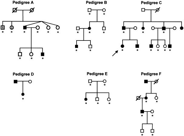
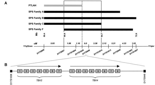
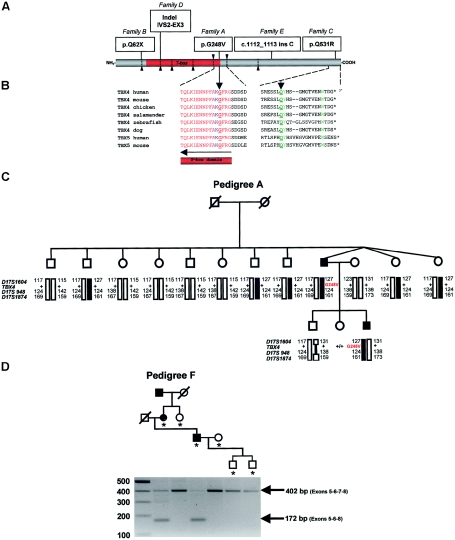
Small patella syndrome (SPS) is an autosomal-dominant skeletal dysplasia characterized by patellar aplasia or hypoplasia and by anomalies of the pelvis and feet, including disrupted ossification of the ischia and inferior pubic rami. We identified an SPS critical region of 5.6 cM on chromosome 17q22 by haplotype analysis. Putative loss-of-function mutations were found in a positional gene encoding T-box protein 4 (TBX4) in six families with SPS. TBX4 encodes a transcription factor with a strongly conserved DNA-binding T-box domain that is known to play a crucial role in lower limb development in chickens and mice. The present identification of heterozygous TBX4 mutations in SPS patients, together with the similar skeletal phenotype of animals lacking Tbx4, establish the importance of TBX4 in the developmental pathways of the lower limbs and the pelvis in humans.


Figures

Figure  1
Figure 1
Characteristic features of the pelvis and the lower limb in individuals with SPS. A, Radiograph of the pelvis of family A's proband at the age of 12 years and 11 mo, showing bilaterally absent ossification of the ischiopubic junction (unblackened arrows), infra-acetabular axe-cut notches (blackened arrows), and elongated femoral necks. B, Radiograph of the pelvis of family C's proband at the age of 19 years and 7 mo. Note that the irregular ossification of the ischiopubic junction (unblackened arrows) and infra-acetabular axe-cut notches (blackened arrows) are less severe, as compared to the pelvic anomalies seen in the proband of family A. C, Radiograph of the knee of family C's proband at the age of 19 years and 7 mo, demonstrating a small patella. D, Feet of an affected male from family C at age 14 years and 8 mo, showing short fourth and fifth rays and an increased space between the first and second toes. (Radiograph A is reproduced from the work of Bongers et al. [2001] with permission from the BMJ Publishing Group.)
Figure  2
Figure 2
Pedigrees of six unrelated kindreds with SPS. Blackened symbols denote individuals with SPS, and unblackened symbols denote unaffected individuals. TBX4 mutation analysis was performed in all individuals indicated by an asterisk (*). The proband of the newly identified Dutch family is indicated by an arrow.
Figure  3
Figure 3
Summary of the linkage analysis and identification of positional candidate genes. A, Linkage analysis of four families with SPS and one family with PTLAH. Microsatellite markers (below the line) span an interval of 24.8 cM at chromosome 17q22. The SPS critical region (D17S957 and D17S1874) is boxed. Linkage analysis of families A and B and the clinical phenotype of families A, B, and F were described elsewhere (Vaněk ; Bongers et al. 2001). Previous linkage studies revealed a PTLAH candidate region at chromosome 17q22 (Mangino et al. 1999). B, Localization and schematic genomic structure of candidate genes TBX2 (exons 1–7) and TBX4 (exons 1–8) (not drawn to scale) (UCSC Genome Bioinformatics).
Figure  4
Figure 4
Molecular analysis of TBX4. A, Distribution of TBX4 gene mutations identified by sequencing. Exon boundaries are identified by arrowheads and dotted lines, and the DNA-binding motif (T-box domain) is colored red. B, Sequence homology analysis of amino acids 248 and 531 and flanking residues (NCBI BLAST). The strongly conserved amino acids of the T-box domain and the C-terminal domain are indicated in red and green colors, respectively. The amino acids that directly contact the DNA are depicted in blue. Glycine 248 and glutamine 531 (amino acids underlined with an arrow) are strongly conserved. C, Segregation of G248V mutation in SPS pedigree A (Bongers et al. 2001). The mutations are present in affected individuals (blackened symbols, G248V) and absent in their healthy siblings (unblackened symbols, +/+). Haplotypes are defined by markers D17S1604, D17S948, and D17S1874 and are encoded by numbers. Blackened bars indicate the risk allele. Note from the mutation and haplotype analysis that the mutation has occurred de novo in the germline of one of the grandparents. D, Segregation of exon 7 skipping in SPS pedigree F (Vaněk, 1981) detected by RT-PCR analysis of the fragment encompassing exons 5–8 of the TBX4 coding region, showing an aberrant sized transcript of 172 bp in the affected individuals on agarose gel. The RT-PCR experiment was performed in all individuals indicated by an asterisk (*).
Figure  5
Figure 5
Protein model of the T-box domain of TBX4, created with the help of the modelling methods described by Chinea et al. (1995) and the resolved structure of TBX3 as template (Coll et al. 2002) (Protein Data Bank). The T-box domains of TBX3 and TBX4 show a high degree of homology (80%) and identity (64%), which ensures a faithful prediction of the TBX4 structure. The 310 helical axis of the T-box domains of TBX3 and TBX4 are 100% identical. The residues of the DNA are represented as a purple stick model. Protein is shown as a ribbon model with red strands, blue helices, and green/turquoise turns and loops. The protein-DNA complex is viewed along the T-box. A, T-box dimer binding to a DNA consensus sequence. Residue G248 (yellow) is located in the C-terminal 310 helix that interacts with the DNA in the minor groove. B, View along the 310 helical axis of the T-box domain. The G248V mutation leads to the introduction of a hydrophobic side chain (yellow atoms in the ball display) that is normally absent. This likely displaces water molecules that stabilize the protein-DNA interaction through hydrogen bonding. Therefore, the G248V mutation is predicted to destabilize the TBX4-DNA interaction. Other highlighted amino acids include F245, K247, and F249 (stick representations).

References

Electronic-Database Information

    1. GenBank, http://www.ncbi.nlm.nih.gov/GenBank/ (for TBX4 [H. sapiens] mRNA [accession number AF188703] and for TBX2 [H. sapiens] mRNA [accession number U28049])
    1. NCBI BLAST, http://www.ncbi.nlm.gov/BLAST/
    1. Online Mendelian Inheritance in Man (OMIM), http://www.ncbi.nlm.nih.gov/Omim/
    1. Protein Data Bank, http://www.rcsb.org/pdb/ (for TBX3 [accession number 1H6F])
    1. UCSC Genome Bioinformatics, http://genome.cse.ucsc.edu/ (for Human Genome Browser)

References

    1. Agarwal P, Wylie JN, Galceran J, Arkhitko O, Li C, Deng C, Grosschedl R, Bruneau BG (2003) Tbx5 is essential for forelimb bud initiation following patterning of the limb field in the mouse embryo. Development 130:623–63310.1242/dev.00191 - DOI - PubMed
    1. Azouz EM, Kozlowski K (1997) Small patella syndrome: a bone dysplasia to recognize and differentiate from the nail-patella syndrome. Pediatr Radiol 27:432–43510.1007/s002470050163 - DOI - PubMed
    1. Bernhang AM, Levine SA (1973) Familial absence of the patella. J Bone Joint Surg Am 55:1088–1090 - PubMed
    1. Bamshad M, Lin R, Law Dj, Watkins WC, Krakowiak PA, Moore ME, Fransceschini P, Lala R, Holmes LB, Gebuhr TC, Bruneau GB, Schinzel A, Seidman JG, Seidman CE, Jorde LB (1997) Mutations in human TBX3 alter limb, apocrine and genital development in ulnar-mammary syndrome. Nat Genet 16:311–315 - PubMed
    1. Basson CT, Bachinsky DR, Lin RC, Levi T, Elkins JA, Soults J, Grayzel D, Kroumpouzou E, Traill T, Leblanc-Straceski J, Renault B, Kucherlapati R, Seidman JG, Seidman CE (1997) Mutations in human TBX5 cause limb and cardiac malformation in Holt-Oram syndrome. Nat Genet 15:30–35 - PubMed

Publication types

MeSH terms

Associated data