. 2004 Apr 15;18(8):901-11.
doi: 10.1101/gad.291004.

The Notch target genes Hey1 and Hey2 are required for embryonic vascular development

Affiliations

The Notch target genes Hey1 and Hey2 are required for embryonic vascular development

Andreas Fischer et al. Genes Dev. .

Abstract

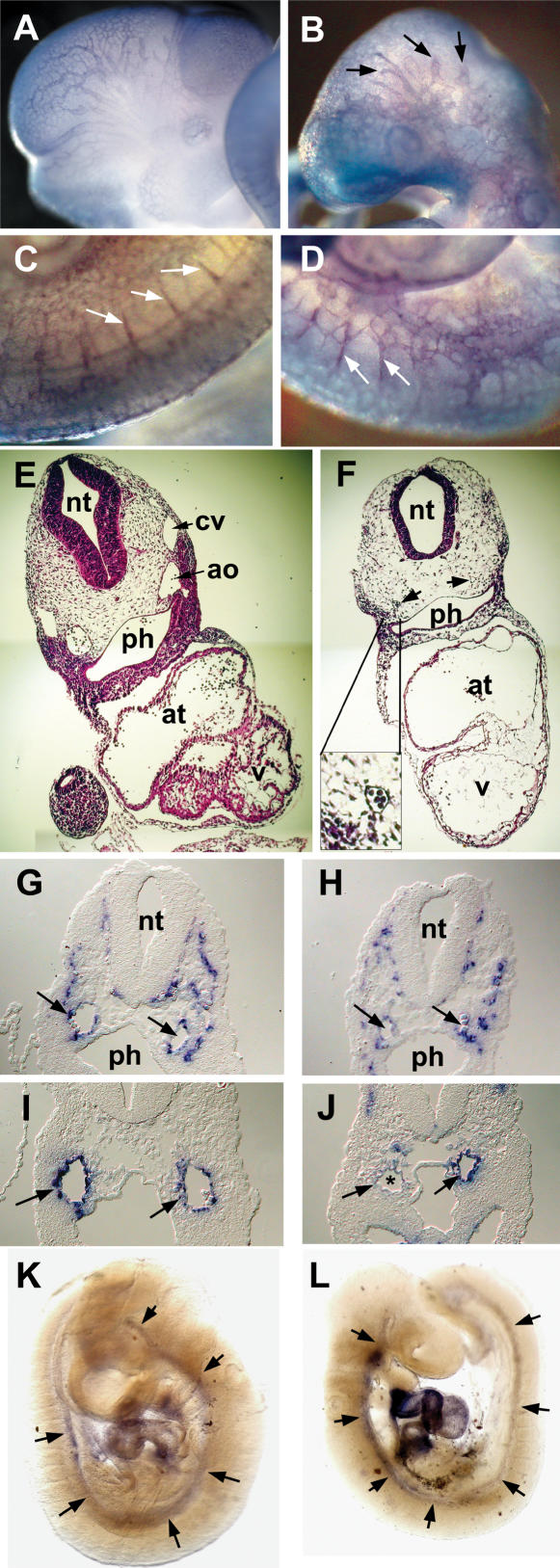
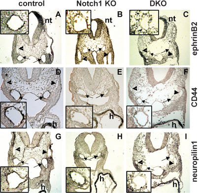
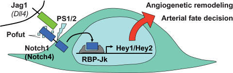
The Delta-Notch signaling pathway plays a central role in the development of most vertebrate organs. The Hey family of bHLH transcription factors are direct targets of Notch signaling. Loss of Hey2 in the mouse leads to cardiac defects with high postnatal lethality. We have now generated a mouse Hey1 knockout that has no apparent phenotypic defect. The combined loss of Hey1 and Hey2, however, results in embryonic death after embryonic day 9.5 (E9.5) with a global lack of vascular remodeling and massive hemorrhage. Initial vasculogenesis appears unaffected, but all subsequently developing major vessels in the embryo and yolk sac are either small or absent. Furthermore, the placental labyrinth completely lacks embryonic blood vessels. Similar vascular defects are observed in Jagged1 and Notch1 knockout mice. In the latter we found Hey1 and Hey2 expression in yolk sacs to be strongly reduced. Remaining large arteries in both Notch1 and Hey1/Hey2 knockout mice fail to express the arterial endothelial markers CD44, neuropilin1, and ephrin-B2. This indicates that Hey1/Hey2 are essential transducers of Notch signals in cardiovascular development that may mediate arterial cell fate decision.


Figures

Figure 1.
Figure 1.
Knockout of the Hey1 locus and embryonic death of Hey1/2 DKO embryos. (A) The targeting construct is depicted above the mouse genomic Hey1 locus containing five exons. Homology regions of 3 kbp (BglII/Ecl136II) and 1.4 kbp (HindIII) containing mainly promoter and 3′untranslated regions, respectively, were used for recombination. The PGK-neo cassette (Neo) is flanked by loxP sites (triangles) for subsequent removal by cre-recombinase. The HSV-TK (TK) cassette was included for negative selection. Flanking genomic probes used for Southern blot hybridization are highlighted by thick lines. (B) BglII; (E) Ecl136II; (H) HindIII; (*) stop codon; (pBS) pBluescript cloning vector backbone. (B) At E10.5, the Hey1/2 DKO embryos can be clearly identified. They are much smaller compared to a het/het littermate (fourth embryo), and they lack prominent mes- and telencephalic vesicles (*). The extensive pericardial sac is highlighted by arrows. (C,D) Already at E9.5, most DKO embryos suffer from massive hemorrhage in the trunk (arrowheads, C) or the pericardial sac (D).
Figure 1.
Figure 1.
Knockout of the Hey1 locus and embryonic death of Hey1/2 DKO embryos. (A) The targeting construct is depicted above the mouse genomic Hey1 locus containing five exons. Homology regions of 3 kbp (BglII/Ecl136II) and 1.4 kbp (HindIII) containing mainly promoter and 3′untranslated regions, respectively, were used for recombination. The PGK-neo cassette (Neo) is flanked by loxP sites (triangles) for subsequent removal by cre-recombinase. The HSV-TK (TK) cassette was included for negative selection. Flanking genomic probes used for Southern blot hybridization are highlighted by thick lines. (B) BglII; (E) Ecl136II; (H) HindIII; (*) stop codon; (pBS) pBluescript cloning vector backbone. (B) At E10.5, the Hey1/2 DKO embryos can be clearly identified. They are much smaller compared to a het/het littermate (fourth embryo), and they lack prominent mes- and telencephalic vesicles (*). The extensive pericardial sac is highlighted by arrows. (C,D) Already at E9.5, most DKO embryos suffer from massive hemorrhage in the trunk (arrowheads, C) or the pericardial sac (D).
Figure 2.
Figure 2.
Placental defects in Hey1/2 DKO mice. A layered structure is visible at E10.5 in both normal (A,C,E,G,I) and Hey1/2 DKO (B,D,F,H) placentas. (A,B) H&E staining shows a close intermingling between maternal blood spaces with small erythrocytes and embryonic vessels with larger, nucleated red blood cells in the control placenta. Especially the labyrinth in the DKO placenta appears cell-rich and devoid of embryonic vessels. (C,D) The giant cell border between the maternal and the embryonic compartment stains positive with a Csh1 (placental lactogen 1) probe. (E,F) Tpbpa (4311) marks the spongiotrophoblast layer that appears unaltered in DKO placentas. (G,H) Vegfr2 (Flk1) stains endothelia of all embryonic vessels in the labyrinth and the chorioallantoic plate. A dotted line highlights the labyrinthine region in DKO mutants, which lacks any staining. (J,K) Fetal vessels only start to invade the trophoblast layer, but fail to branch and extend any further as seen by H&E (J) and endothelial Vegfr2 (K) staining. Abortive buds are marked by triangles. (I) In a control placenta, the labyrinthine layer is characterized by intermingling and close apposition of maternal and embryonic blood spaces to facilitate nutrient and gas exchange. (L,M) Both Hey1 and Hey2 are expressed in endothelia of embryonic vessels in the chorioallantoic plate (arrowheads) of controls and faintly if at all in the embryonic labyrinth. (gc) Giant cells; (sp) spongiotrophoblast; (la) labyrinth; (cp) chorionic plate; (al) allantois.
Figure 3.
Figure 3.
Yolk sac defects in Hey1/2 DKO mice. There is a clear difference between intact het/het controls (A,C) and DKO (B,D) yolk sacs. (A,B) At E9.5, a vascular network with embryonic red blood cells can be seen. In A, large vessels are clearly visible, whereas DKO embryos only exhibit the primitive vascular network without further reorganization. (C,D) At E10.5, large vitelline vessels are present in controls, but in DKO embryos the yolk sacs are pale and begin to degenerate. (E,F) Sections of E9.5 yolk sacs clearly show that initial vasculogenesis is comparable in het/het control (E) and DKO (F) embryos. (G,H) The presence of blood cells in sections and intact yolk sacs suggests that hematopoiesis from blood islands is not affected. PECAM staining of blood vessel endothelia confirms highly organized vascular branches in controls (G), but complete absence of remodeling in yolk sacs from a DKO littermate (H).
Figure 4.
Figure 4.
Vascular defects in Hey1/2 DKO embryos. (A–D) Comparison of whole-mount PECAM antibody staining of E10.5 control (A,C) and DKO (B,D) embryos reveals intact vasculogenesis. Large cranial vessels appear truncated in mutants (arrows). Angiogenetic sprouts of intersomitic vessels (white arrows) are present, but the vascular pattern in the trunk is rather coarse. (E,F) H&E-stained cross-sections revealed reduction or loss of aorta (arrows) and cardinal vein. The myocardial wall is thinner in mutants, and ventricular trabeculation is missing. The neural tube is thinner and the mesenchymal compartment is cell-poor in mutants. (G,H) In situ hybridization with the endothelial marker VE-Cadherin identifies the aorta (arrows) and multiple smaller vessels. (I,J) The smooth muscle cell marker SM22 stains the aorta in controls (I), but in DKO embryos (J) staining is variable with partial or even complete loss (*) of the hybridization signal. (K,L) Whole-mount in situ hybridization for SM22 shows that the aorta is associated with smooth muscle cells along its length in controls and mutants, at least at this level of resolution. (ao) Aorta; (at) atrium; (cv) cardinal vein; (nt) neural tube; (ph) pharynx; (v) ventricle.
Figure 5.
Figure 5.
Lack of vascular ephrin-B2, CD44, and neuropilin1 expression in Notch1 and Hey1/2 KO embryos. Expression of the arterial endothelial markers ephrin-B2 (AC), CD44 (DF), and neuropilin1 (GI) in wild-type (A,D,G), Notch1–/– (B,E,H), and Hey1/2 DKO (C,F,I) embryos at E9.5. Immunohistochemistry of transverse sections reveals staining of both aortae (arrows) in control embryos, whereas the cardinal veins (arrowheads) are negative. Similar sections of the mutant embryos still exhibit cardiac CD44 and neuropilin1 expression as well as ephrin-B2 staining in the neural tube, but the aortae are clearly not stained. (h) Heart; (nt) neural tube. Enlarged views of an aortic vessel for each case are shown in insets.
Figure 6.
Figure 6.
Notch signal transduction in endothelial biology. KO studies in the mouse identified a complete scenario for Notch signal transduction in arterial endothelial cells. The Jag1 (and presumably also the Dll4) ligand appear critical for endothelial cells and their precursors. Notch1, aided by Notch4, transmits the signal, dependent on it own glycosylation and cleavage by Pofut and Ps1/2. The Notch intracellular domain together with RBP-Jk activates Hey1 and Hey2 transcription. Knockout of any of these components leads to very similar vascular deficiencies.

References

    1. Adams, R.H. 2003. Molecular control of arterial-venous blood vessel identity. J. Anat. 202: 105–112. - PMC - PubMed
    1. Batlle, E., Henderson, J.T., Beghtel, H., van den Born, M.M., Sancho, E., Huls, G., Meeldijk, J., Robertson, J., van de Wetering, M., Pawson, T., et al. 2002. Beta-catenin and TCF mediate cell positioning in the intestinal epithelium by controlling the expression of EphB/ephrinB. Cell 111: 251–263. - PubMed
    1. Carmeliet, P. 2003. Angiogenesis in health and disease. Nat. Med. 9: 653–660. - PubMed
    1. Chi, J.T., Chang, H.Y., Haraldsen, G., Jahnsen, F.L., Troyanskaya, O.G., Chang, D.S., Wang, Z., Rockson, S.G., van de Rijn, M., Botstein, D., et al. 2003. Endothelial cell diversity revealed by global expression profiling. Proc. Natl. Acad. Sci. 100: 10623–10628. - PMC - PubMed
    1. Donovan, J., Kordylewska, A., Jan, Y.N., and Utset, M.F. 2002. Tetralogy of fallot and other congenital heart defects in Hey2 mutant mice. Curr. Biol. 12: 1605–1610. - PubMed

Publication types