. 2004 Apr 15;18(8):937-51.
doi: 10.1101/gad.1190304.

Hedgehog signaling in the neural crest cells regulates the patterning and growth of facial primordia

Affiliations

Hedgehog signaling in the neural crest cells regulates the patterning and growth of facial primordia

Juhee Jeong et al. Genes Dev. .

Abstract

Facial abnormalities in human SHH mutants have implicated the Hedgehog (Hh) pathway in craniofacial development, but early defects in mouse Shh mutants have precluded the experimental analysis of this phenotype. Here, we removed Hh-responsiveness specifically in neural crest cells (NCCs), the multipotent cell type that gives rise to much of the skeleton and connective tissue of the head. In these mutants, many of the NCC-derived skeletal and nonskeletal components are missing, but the NCC-derived neuronal cell types are unaffected. Although the initial formation of branchial arches (BAs) is normal, expression of several Fox genes, specific targets of Hh signaling in cranial NCCs, is lost in the mutant. The spatially restricted expression of Fox genes suggests that they may play an important role in BA patterning. Removing Hh signaling in NCCs also leads to increased apoptosis and decreased cell proliferation in the BAs, which results in facial truncation that is evident by embryonic day 11.5 (E11.5). Together, our results demonstrate that Hh signaling in NCCs is essential for normal patterning and growth of the face. Further, our analysis of Shh-Fox gene regulatory interactions leads us to propose that Fox genes mediate the action of Shh in facial development.


Figures

Figure 1.
Figure 1.
Expression of Shh and Ptch1 in relation to facial development in the mouse embryo. Lateral views (A,E,I) and frontal views (B,F,J) of whole-mount β-galactosidase staining of heads from ShhlacZ/+ embryos. Lateral views (C,G,K) and frontal views (D,H,L) of whole-mount β-galactosidase staining of heads from Ptch1lacZ/+ embryos. (MX) Transverse sections through the heads of ShhlacZ/+ (M,P,S,V), Ptch1lacZ/+ (N,Q,T,W), and Wnt1-Cre;R26R-LacZ (O,R,U,X) embryos. Stages of development as indicated in each panel. Bar, 0.5 mm.
Figure 2.
Figure 2.
Craniofacial phenotypes of Wnt1-Cre;Smon/c embryos. (AF) Heads of wild-type (A,C,E) and Wnt1-Cre;Smon/c (B,D,F) embryos at the stages indicated. (G,H) Whole-mount β-galactosidase staining of heads from Wnt1-Cre;R26R-LacZ (G) and Wnt1-Cre;Smon/c;R26R-LacZ (H) embryos at E10.5. (I,J) Facial ganglia labeled by whole-mount immunostaining for neurofilament in wild-type (I) and Wnt1-Cre;Smon/c (J) embryos at E10.5. (KU) Skeletal preparations of heads from wild-type (K,M,O,R,T) and Wnt1-Cre;Smon/c (L,N,P,Q,S,U) embryos at E18.5. (K,L) Lateral views. (M,N) Skull bases with dentaries removed. (OQ) Dentaries. In Q,the inner and outer layers of the mutant dentary have been separated to show Meckel's cartilage. (R,S) Otic capsules and the associated elements. (T,U) Hyoid and laryngeal cartilages. (VX) Schematics of head skeleton. (W) Green indicates skeletal elements of NCC origin; blue indicates skeletal elements of mesodermal origin. (X) Pink indicates mildly reduced or malformed in Wnt1-Cre;Smon/c embryos; red indicates severely reduced in Wnt1-Cre;Smon/c embryos; black indicates absent in Wnt1-Cre;Smon/c embryos. (AC) Arytenoid cartilage; (AP) angular process; (AS) alisphenoid; (BH) basihyoid; (BO) basioccipital; (BS) basisphenoid; (CC) cricoid cartilage; (CDP) condylar process; (CH) ceratohyoid; (CRP) coronoid process; (DNT) dentary; (EO) exoccipital; (FN) frontal; (GN) gonial; (Il) lower incisor; (IN) incus; (IP) interparietal; (Iu) upper incisor; (JG) jugal; (LA) lacrimal; (MA) malleus; (MC) Meckel's cartilage; (ME) mesethmoid; (Mo) molar; (MX) maxilla; (MXPP) palatal process of maxilla; (MXZP) zygomatic process of maxilla; (NA) nasal bone; (NC) nasal cartilage; (NF) neurofilament; (OC) otic capsule; (OS) orbitosphenoid; (PA) parietal; (PL) palatine; (PM) premaxilla; (PS) presphenoid; (PT) pterygoid; (SO) supraoccipital; (SP) styloid process; (SQ) squamosal; (ST) stapes; (TC) thyroid cartilage; (TH) thyrohyoid; (TR) tracheal rings; (TY) tympanic ring; (VM) vomer. Bar, 1 mm. VX were modified with permission from Depew et al. (1999). © 1999 The Company of Biologists Limited.
Figure 5.
Figure 5.
Expression of non-Fox family facial markers in E10.5 embryos. Frontal views of whole-mount in situ hybridization samples. Genotypes of the embryos and probes as indicated in each panel. Bar, 0.25 mm.
Figure 3.
Figure 3.
Expression of Fox genes in the facial primordia of E9.5 embryos. Lateral views (odd-number columns) and frontal views (even-number columns) of whole-mount in situ hybridization samples. Genotypes of the embryos and probes as indicated in each panel.
Figure 4.
Figure 4.
Expression of Fox genes in the facial primordia of E10.5 embryos. Lateral views (odd-number columns) and frontal views (even-number columns) of whole-mount in situ hybridization samples. Genotypes of the embryos and probes as indicated in each panel.
Figure 8.
Figure 8.
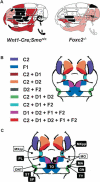
A model of BA patterning by the Shh–Fox genetic pathway. (A) Summary of head skeleton defects of Wnt1-Cre;Smon/c and Foxc2–/– embryos at E18.5. The phenotype of Foxc2–/– is based on Winnier et al. (1997), which reported more severe defects than Iida et al. (1997). Pink indicates mildly reduced or malformed; red indicates severely reduced; black indicates absent. (B) Summary of the expression patterns of Foxc2, Foxd1, Foxd2, Foxf1, and Foxf2 in wild-type facial primordia at E10.5. (C) The “Fox code map” from B, superimposed with the putative fate map of the first and second BAs. The face is symmetric around the midline, although each element is labeled on one side only due to space limitations. (DNT) Dentary; (GN) gonial; (Il) lower incisor; (MA) malleus; (MO) molar; (MXpp) palatal process of maxilla; (MXzp) zygomatic process of maxilla; (PL) palatine; (SP) styloid process; (ST) stapes; (TO) tongue; (TY) tympanic ring. White characters in black box label the elements absent in Wnt1-Cre;Smon/c embryos, whereas the black characters in white box label those present in the mutant. A was modified with permission from Depew et al. (1999). © 1999 The Company of Biologists Limited.
Figure 6.
Figure 6.
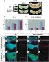
Growth defects in the MNAs of Wnt1-Cre;Smon/c embryos. (A) MNAs from wild-type (top) and Wnt1-Cre;Smon/c (bottom) embryos at the stages indicated. (B,C) Quantification of mediolateral (B) and dorsoventral (C) development of the wild-type and Wnt1-Cre;Smon/c MNAs. Three samples of each genotype were measured at each stage. (DO) Immunofluorescence with antibodies against cleaved-caspase-3 (red signal; DK) and phospho-histone H3 (red signal; LO) to detect apoptotic and mitotic cells, respectively. (D,E,H,I,L,M) Transverse sections through the MNAs of Wnt1-Cre;R26R-YFP embryos. (F,G,J,K,N,O) Transverse sections through the MNAs of Wnt1-Cre;Smon/c;R26R-YFP embryos. Stages of development as indicated. Green signal indicates YFP from R26R-YFP; blue signal indicates Topro3 counterstain for nuclei. (D,H,L,F,J,N) Merged images of red, green, and blue channels. (E,I,M,G,K,O) Red channel only. Bar, 0.25 mm.
Figure 7.
Figure 7.
Loss of tongue and lower incisors in Wnt1-Cre; Smon/c embryos. (A,B) Coronal sections through heads of Wnt1-Cre;R26R-YFP (A) and Wnt1-Cre;Smon/c;R26R-YFP (B) embryos at E12.5. Green signal indicates YFP from R26R-YFP; blue signal indicates Topro3 counterstain for nuclei. (CH) Whole-mount in situ hybridization analyses of tongue and tooth development in wild-type (C,E,G) and Wnt1-Cre;Smon/c (D,F,H) embryos. (C,D) Frontal views of Myf5 expression at E10.75. (E,F) Dorsal views of Shh expression in the MNA at E11.5. (G,H) Frontal views of Pax9 expression at E11.5. (IN) Histological analysis of teeth in coronal sections through heads of wild-type (I,K,M) and Wnt1-Cre;Smon/c (J,L,N) embryos at E18.5. (de) Dental ectoderm; (ep) tongue epithelium; (il) lower incisor; (iu) upper incisor; (mes) tongue mesenchyme; (ml) lower molar; (mu) upper molar; (mus) tongue muscle; (to) tongue. Bar, 0.25 mm.

References

    1. Ahlgren, S.C. and Bronner-Fraser, M. 1999. Inhibition of Sonic hedgehog signaling in vivo results in craniofacial neural crest cell death. Curr. Biol. 9: 1304–1314. - PubMed
    1. Barlow, A.J. and Francis-West, P.H. 1997. Ectopic application of recombinant BMP-2 and BMP-4 can change patterning of developing chick facial primordia. Development 124: 391–398. - PubMed
    1. Beverdam, A., Merlo, G.R., Paleari, L., Mantero, S., Genova, F., Barbieri, O., Janvier, P., and Levi, G. 2002. Jaw transformation with gain of symmetry after Dlx5/Dlx6 inactivation: Mirror of the past? Genesis 34: 221–227. - PubMed
    1. Carlsson, P. and Mahlapuu, M. 2002. Forkhead transcription factors: Key players in development and metabolism. Dev. Biol. 250: 1–23. - PubMed
    1. Chai, Y., Jiang, X., Ito, Y., Bringas, P., Han, J., Rowitch, D.H., Soriano, P., McMahon, A.P., and Sucov, H.M. 2000. Fate of the mammalian cranial neural crest during tooth and mandibular morphogenesis. Development 127: 1671–1679. - PubMed

Publication types

MeSH terms