. 2004 Apr 15;18(8):952-63.
doi: 10.1101/gad.1174704.

Runx2 and Runx3 are essential for chondrocyte maturation, and Runx2 regulates limb growth through induction of Indian hedgehog

Affiliations

Runx2 and Runx3 are essential for chondrocyte maturation, and Runx2 regulates limb growth through induction of Indian hedgehog

Carolina A Yoshida et al. Genes Dev. .

Abstract

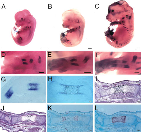
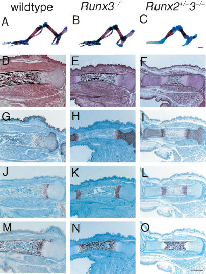
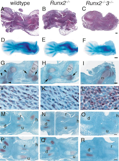
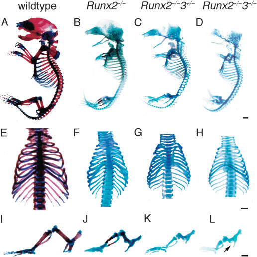
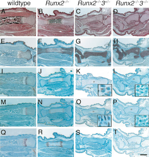
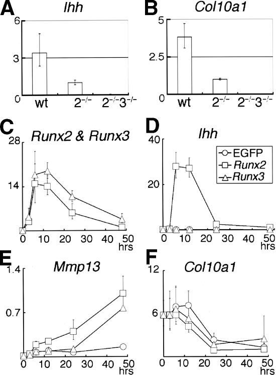
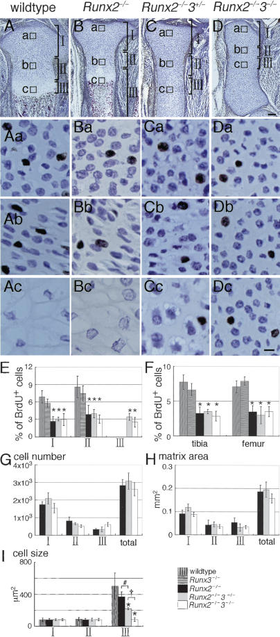
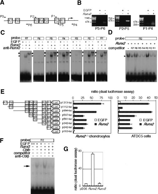
The differentiation of mesenchymal cells into chondrocytes and chondrocyte proliferation and maturation are fundamental steps in skeletal development. Runx2 is essential for osteoblast differentiation and is involved in chondrocyte maturation. Although chondrocyte maturation is delayed in Runx2-deficient (Runx2(-/-)) mice, terminal differentiation of chondrocytes does occur, indicating that additional factors are involved in chondrocyte maturation. We investigated the involvement of Runx3 in chondrocyte differentiation by generating Runx2-and-Runx3-deficient (Runx2(-/-)3(-/-)) mice. We found that chondrocyte differentiation was inhibited depending on the dosages of Runx2 and Runx3, and Runx2(-/-)3(-/-) mice showed a complete absence of chondrocyte maturation. Further, the length of the limbs was reduced depending on the dosages of Runx2 and Runx3, due to reduced and disorganized chondrocyte proliferation and reduced cell size in the diaphyses. Runx2(-/-)3(-/-) mice did not express Ihh, which regulates chondrocyte proliferation and maturation. Adenoviral introduction of Runx2 in Runx2(-/-) chondrocyte cultures strongly induced Ihh expression. Moreover, Runx2 directly bound to the promoter region of the Ihh gene and strongly induced expression of the reporter gene driven by the Ihh promoter. These findings demonstrate that Runx2 and Runx3 are essential for chondrocyte maturation and that Runx2 regulates limb growth by organizing chondrocyte maturation and proliferation through the induction of Ihh expression.


Figures

Figure 1.
Figure 1.
Runx3 expression in Runx3+/– mice and skeletal development in Runx3–/– mice during embryogenesis. (AH), Runx3 expression in Runx3+/– mice at E12.5 (A,D,G), E13.5 (B,E), E15.5 (C,F) and E16.5 (H). We examined Runx3 expression by the activity of β-galactosidase, whose gene had been integrated in exon 3 of Runx3, in Runx3+/– mice. (AF) Runx3 was expressed in cartilaginous skeletons. (G,H) Runx3 was specifically expressed in chondrocytes but not in osteoblasts in the skeleton, and its expression was up-regulated in prehypertrophic chondrocytes and down-regulated in terminal hypertrophic chondrocytes. (IL) Skeletal development in Runx3–/– mice at E15.5. H&E and Kossa staining (I,J) and in situ hybridization using Col10a1 antisense probe (K,L) of wild-type (I,K) and Runx3–/– (J,L) tibiae. Calcification was stained black in I and J. We detected no signal using a sense probe of Col10a1 (data not shown). The diaphyses of wild-type tibiae were composed of calcified cartilage that was negative for Col10a1 expression (K), and blood vessels had invaded the diaphyses (I). (J,L) The diaphyses of Runx3–/– tibiae were composed of Col10a1-positive hypertrophic chondrocytes without vascular invasion. Bars: AC, 1 mm; DF, 500 μm; GL, 100 μm.
Figure 2.
Figure 2.
Skeletal development in Runx3–/– and Runx2+/–3–/– mice. (AC) Hind limb skeletons of wild-type (A), Runx3–/– (B), and Runx2+/–3–/– (C) newborns. Calcified tissues were stained red with Alizarin red, and cartilage was stained blue with Alcian blue. Wild-type and Runx3–/– limb skeletons showed a similar pattern of mineralization (A,B), whereas the mineralized regions of Runx2+/–3–/– limbs were much smaller (C). (DF) Histological analysis of the tibiae of wild-type (D), Runx3–/– (E), and Runx2+/–3–/– (F) embryos at E18.5. The sections were stained with H&E and Kossa. (GO) In situ hybridization using the antisense probes of Col2a1 (GI), Col10a1 (JL), and osteopontin (MO). We detected no signal using sense probes of Col2a1, Col10a1, or osteopontin (data not shown). In the tibiae of wild-type and Runx3–/– embryos, the cartilage was largely replaced by bone (D,E), osteopontin-positive osteoblasts occupied the bone marrow (M,N), and Col2a1-positive (G,H) or Col10a1-positive (J,K) chondrocytes were observed in the epiphyses. Runx2+/–3–/– tibiae were still cartilaginous (F), and the chondrocytes expressed Col2a1 (I) or Col10a1 (L) except for those in the mineralized diaphyses, which were occupied by osteopontin-positive terminal hypertrophic chondrocytes (O). Bars: AC, 1 mm; DO, 500 μm.
Figure 3.
Figure 3.
Early skeletal development in wild-type, Runx2–/–, and Runx2–/–3–/– embryos and in situ hybridization for Sox9 and Ihh expression. We used the forelimbs (AC,GR) and hind limbs (DF) of wild-type (A,D,G,J,M,P), Runx2–/– (B,E,H,K,N,Q), and Runx2–/–3–/– (C,F,I,L,O,R) embryos for the analyses. (AC) H&E staining at E12.5. (DF) Whole-mount Alcian blue staining at E13.5. (GL) In situ hybridization using Sox9 antisense probe at E13.5. (JL) Higher magnification of boxed regions in GI, respectively. (MR) In situ hybridization using Ihh antisense probe at E12.5 (MO) and E13.5 (P–R). We detected no signal using the sense probe of Sox9 and Ihh (data not shown). Cartilaginous anlagen was clearly seen in the wild-type (A) and Runx2–/– (B) embryos but not in the Runx2–/–3–/– embryos (C) at E12.5. Alcian blue staining of the hind limbs of E13.5 Runx2–/–3–/– embryos (F) was weak, compared with that of the wild-type (D) and Runx2–/– (E) embryos. (G,H,J,K) In the forelimb skeletons of wild-type and Runx2–/– embryos, the peripheral regions of the epiphyses of the humeri, radii, and ulnae (arrows in G,H), and most regions of the carpal and metacarpal bones (arrowheads in G) were composed of immature chondrocytes that strongly expressed Sox9, and the remaining regions of the humeri, radii, and ulnae, which were composed of differentiating chondrocytes, weakly expressed Sox9. (I,L) In the Runx2–/–3–/– forelimb skeletons, all of the chondrocytes strongly expressed Sox9. Ihh was strongly detected in immature chondrocytes. especially in digits of wild-type, Runx2–/–, and Runx2–/–3–/– mice. (h) humerus; (r) radius; (u) ulna; (d) digit. Bars: A–C,GI,MR, 100 μm; DF, 500 μm; JL, 10 μm.
Figure 4.
Figure 4.
Examination of the skeletal system of wild-type, Runx2–/–, Runx2–/–3+/–, and Runx2–/–3–/– mice at the newborn stage. Skeletons of wild-type (A,E,I), Runx2–/– (B,F,J), Runx2–/–3+/– (C,G,K), and Runx2–/–3–/– (D,H,L) newborns. (AD) Whole skeletons. (EH) Chest walls. (IL) Hind limbs. Although mineralization was observed in restricted skeletal parts including the limbs, vertebrae, and ribs of Runx2–/– mice (B,F,J), it was completely absent in Runx2–/–3+/– (C,G,K) and Runx2–/–3–/– mice (D,H,L). Note that the limb length was reduced depending on the dosages of Runx2 and Runx3, and that Runx2–/–3–/– mice have severely shortened but relatively thick limbs (IL). Runx2–/–3–/– mice completely lack a pubic bone structure (arrow in L). Bars: AL, 1 mm.
Figure 5.
Figure 5.
Histological examination and in situ hybridization for Col2a1, Pthr1, Ihh, and Col10a1 in the tibiae of wild-type, Runx2–/–, Runx2–/–3+/–, and Runx2–/–3–/– embryos at E18.5. Sections of tibiae from wild-type (A,E,I,M,Q), Runx2–/– (B,F,J,N,R), Runx2–/–3+/– (C,G,K,O,S), and Runx2–/–3–/– (D,H,L,P,T) embryos were examined by H&E and Kossa staining (AD) and in situ hybridization using the antisense probes of Col2a1 (EH), Pthr1 (IL), Ihh (M–P), or Col10a1 (QT). The boxed regions in K, L, O, and P are magnified in the respective insets. On in situ hybridization of Pthr1 and Ihh, signal detection using the sections from Runx2–/– and Runx2–/–3+/– tibiae always took a longer period of time than that using sections from wild-type tibiae. Further, we failed to detect Pthr1 and Ihh expression in Runx2–/–3–/– tibiae even upon prolonged incubation. We detected no signal using sense probes of Col2a1, Pthr1, Ihh, and Col10a1 (data not shown). In the tibiae of wild-type embryos, the cartilage was largely replaced by bone, whereas all of the Runx2–/–, Runx2–/–3+/–, and Runx2–/–3–/– tibiae were cartilaginous. In the Runx2–/– tibiae, the diaphyses were composed of calcified cartilage and chondrocytes in the metaphyses expressed Pthr1, Ihh, or Col10a1. In contrast, Runx2–/– 3+/– and Runx2–/–3–/– tibiae showed no mineralization and the chondrocytes in the entire tibiae expressed Col2a1 but not Col10a1. Pthr1 and Ihh expression were very weakly detected in the diaphyses of Runx2–/–3+/– tibiae, but they were not detected in Runx2–/–3–/– tibiae (see insets in K,L,O,P). Bar, 500 μm.
Figure 7.
Figure 7.
Regulation of Ihh expression by Runx2 and Runx3. (A,B) Real-time RT–PCR analysis of Ihh (A) and Col10a1 (B) expression using RNA extracted from wild-type (wt), Runx2–/– (2–/–), and Runx2–/–3–/– (2–/–3–/–) limbs at E18.5. The mean of two to three embryos is shown. The values in Runx2–/– limbs were defined as 1, and relative values are shown. (CF) Real-time RT–PCR analysis of Runx2–/– chondrocyte cultures. Runx2–/– chondrocytes were infected with EGFP-expressing (○), Runx2-and-EGFP-expressing (□), or Runx3-and-EGFP-expressing (▵) adenovirus. Infected cells were harvested at the indicated times after the onset of viral infection, and Runx2 (C), Runx3 (C), Ihh (D), Mmp13 (E), and Col10a1 (F) expression were examined by real-time RT–PCR. The value of Runx2 mRNA expression at 48 h was defined as 1, and the relative values of Runx2 and Runx3 mRNA expression are shown in the same graph (Runx2 and Runx3). In the cells infected with EGFP-expressing adenovirus, the values of Runx2 mRNA expression were nearly zero and the values of Runx3 mRNA expression were very low; therefore, they were not included in the graph. The value of Ihh, Mmp13, or Col10a1 mRNA expression in Runx2-and-EGFP-expressing adenovirus infection at 48 h was defined as 1, and relative values are shown. Adenoviral introduction of Runx2 strongly up-regulated Ihh expression, whereas adenoviral introduction of Runx3 did not up-regulate Ihh expression. In the cells infected with any of the three adenoviruses, the level of Col10a1 expression was at the background level during the culture period examined. Data represent the mean of three to six wells, and representative data of three independent experiments are shown.
Figure 6.
Figure 6.
Analysis of chondrocyte proliferation in the limbs by BrdU labeling. (AD) Immunohistochemistry of tibiae from BrdU-labeled wild-type (A), Runx2–/– (B), Runx2–/–3+/– (C), and Runx2–/– 3–/– (D) embryos at E18.5 using anti-BrdU antibody. The sections were counterstained with toluidine blue. Magnified views of the boxed regions (a,b,c) are shown in the same columns. (A,B) In wild-type and Runx2–/– tibiae, the boxed regions a, b, and c represent resting, proliferating, and hypertrophic chondrocytes, respectively. (C,D) In the Runx2–/–3+/– and Runx2–/–3–/– tibiae, the boxed regions a, b, and c represent chondrocytes in the epiphyses, metaphyses, and diaphyses, respectively. The growth plates were well formed in wild-type tibiae (A) and in Runx2–/– tibiae (B) but not in Runx2–/– 3+/– tibiae (C) or Runx2–/–3–/– tibiae (D). (AbDb) The columnar alignment of chondrocytes, which is seen in the layer of proliferating chondrocytes, is well formed in the wild-type and Runx2–/– tibiae, deformed in the Runx2–/–3+/– tibiae, and completely absent in the Runx2–/–3–/– tibiae. (C,D) The diaphyses of Runx2–/–3+/– tibiae were composed of slightly enlarged chondrocytes, whereas the entire tibiae of Runx2–/–3–/– embryos were composed of homogeneously small chondrocytes. (EI) Measurement of the frequency of BrdU-positive cells (E,F), cell number (G), matrix area (H), and cell size (I) using the tibiae of six wild-type embryos, two Runx3–/– embryos, six Runx2–/– embryos, one Runx2–/–3+/– embryo, and two Runx2–/–3–/– embryos. We measured these parameters in each region shown in AD. (A,B) In wild-type and Runx2–/– tibiae, the regions I, II, and III represent resting, proliferating, and hypertrophic and terminal hypertrophic chondrocytes, respectively. (C,D) In the Runx2–/–3+/– and Runx2–/–3–/– tibiae, the regions I, II, and III were arbitrarily determined in the proximal half of the tibiae and represent chondrocytes in the epiphysial, metaphysical, and diaphysial parts of the tibiae, respectively. (F) We also counted the number of BrdU-positive cells in femurs, because chondrocyte maturation in Runx2–/– femurs is more severely inhibited than that in Runx2–/– tibiae (Inada et al. 1999). (F) In the measurement of BrdU-positive cells in whole tibiae and femurs, we counted the number of BrdU-positive cells and the total number of cells in regions I and II in wild-type and Runx2–/– mice and in regions I, II, and III in Runx2–/– 3+/– and Runx2–/–3–/– mice, and the mean of the percentage of BrdU-positive cells ± standard deviation (std. dev.) is shown. We measured all of the parameters in three sections of each bone, and the mean ± std. dev. is shown. (*) p < 0.0001 compared with wild-type mice; (#) p < 0.0001; (†) p < 0.0001 as determined by one-way ANOVA. Bars: AD, 100 μm; AaDc, 10 μm.
Figure 8.
Figure 8.
Regulation of the Ihh promoter by Runx2. (A) Schematic presentation of seven putative Runx-binding sites (R1–R7) and primers used for ChIP in the promoter region of the mouse Ihh gene. (B) ChIP. Runx2–/– chondrocytes were infected with EGFP- or Runx2-and-EGFP-expressing adenovirus. After sonication, the cell lysates were immunoprecipitated by anti-Runx2 antibody, and the binding of Runx2 to the Ihh promoter region was examined by PCR using the primers shown in A. Bands of correct sizes were detected in Runx2-expressing cells, and the amplified DNA contained each Ihh promoter fragment, which was confirmed by sequencing (data not shown). (C) Binding of Runx2 to the putative Runx binding sequences. Runx2–/– chondrocytes were infected with EGFP- or Runx2-and-EGFP-expressing adenovirus, and EMSA was performed using each of the R1–R7 probes. Runx2 strongly bound to R1, R2, R4, and R5, less strongly to R7, and weakly to R3 and R6. All of the specific bands (arrowheads) were super-shifted with anti-Runx2 antibody (*). All of the specific bands were competed with the respective unlabeled oligonucleotide (data not shown). (D) The affinity of Runx2 for each putative Runx binding sequence. Runx2–/– chondrocytes were infected with Runx2-and-EGFP-expressing adenovirus, and EMSA was performed using labeled R2 probe in the presence of each unlabeled oligonucleotide (R1–R7/×200). The formation of the Runx2–R2 complex was strongly inhibited in the presence of unlabeled R1, R2, R4, or R5 oligonucleotides, less strongly inhibited in the presence of unlabeled R7 oligonucleotides, but barely inhibited in the presence of unlabeled R3 or R6 oligonucleotides. (E) Reporter assays of the Ihh promoter. The reporter plasmid containing each Ihh promoter construct and pRL-CMV vector were cotransfected into Runx2–/– chondrocytes (left) or ATDC5 cells (right). After culture for 24 h, Runx2 was adenovirally introduced into the cells at an MOI of 10. The data show the relative level of luciferase activity against the level of Renilla luciferase activity. The values are mean ± S.E. of four wells, and representative data from four independent experiments are shown. (F) Binding of Runx3 to the R2 oligonucleotides. Runx2–/– chondrocytes were infected with retrovirus expressing EGFP alone or Cbfb and EGFP. After 1 d, the cells were infected with EGFP- or Runx3-and-EGFP-expressing adenovirus, and EMSA was performed using the R2 probe. The introduction of Runx3 alone showed a specific band (arrow), and the intensity of the specific band was increased by the introduction of Cbfb. The specific band was competed with unlabeled R2 oligonucleotides (×200). Addition of antibody against Cbfβ prevented formation of the specific Runx3–Cbfβ–DNA complexes. (G) Reporter assay using the 1.3-kb Ihh promoter. To compare the capacities of Runx2 and Runx3 in the transcriptional activation of Ihh promoter, Runx2–/– chondrocytes were transiently transfected with DNA mixture containing p1313-luc, Runx2- or Runx3-expressing vector or empty pSG5, and pRL-CMV. The luciferase activity was normalized to Renilla luciferase activity.

References

    1. Akiyama, H., Chaboissier, M.C., Martin, J.F., Schedl, A., and de Crombrugghe, B. 2002. The transcription factor Sox9 has essential roles in successive steps of the chondrocyte differentiation pathway and is required for expression of Sox5 and Sox6. Genes & Dev. 16: 2813–2828. - PMC - PubMed
    1. Bae, S.C., Takahashi, E., Zhang, Y.W., Ogawa, E., Shigesada, K., Namba, Y., Satake, M., and Ito, Y. 1995. Cloning, mapping and expression of PEBP2 αC, a third gene encoding the mammalian Runt domain. Gene 159: 245–248. - PubMed
    1. Bi, W., Deng, J.M., Zhang, Z., Behringer, R.R., and de Crombrugghe, B. 1999. Sox9 is required for cartilage formation. Nat. Genet. 22: 85–89. - PubMed
    1. Bitgood, M.J. and McMahon, A.P. 1995. Hedgehog and Bmp genes are coexpressed at many diverse sites of cell–cell interaction in the mouse embryo. Dev. Biol. 172: 126–138. - PubMed
    1. Enomoto, H., Enomoto-Iwamoto, M., Iwamoto, M., Nomura, S., Himeno, M., Kitamura, Y., Kishimoto, T., and Komori, T. 2000. Cbfa1 is a positive regulatory factor in chondrocyte maturation. J. Biol. Chem. 275: 8695–8702. - PubMed

Publication types