. 2004 May 4;101(18):6876-81.
doi: 10.1073/pnas.0307306101. Epub 2004 Apr 23.

Infertility with defective spermatogenesis and hypotestosteronemia in male mice lacking the androgen receptor in Sertoli cells

Affiliations

Infertility with defective spermatogenesis and hypotestosteronemia in male mice lacking the androgen receptor in Sertoli cells

Chawnshang Chang et al. Proc Natl Acad Sci U S A. .

Abstract

Androgens and the androgen receptor (AR) play important roles in male fertility, although the detailed mechanisms, particularly how androgen/AR influences spermatogenesis in particular cell types, remain unclear. Using a Cre-Lox conditional knockout strategy, we generated a tissue-specific knockout mouse with the AR gene deleted only in Sertoli cells (S-AR(-/y)). Phenotype analyses show the S-AR(-/y) mice were indistinguishable from WT AR mice (B6 AR(+/y)) with the exception of testes, which were significantly atrophied. S-AR(-/y) mice were infertile, with spermatogenic arrest predominately at the diplotene premeiotic stage and almost no sperm detected in the epididymides. S-AR(-/y) mice also have lower serum testosterone concentrations and higher serum leuteinizing hormone concentrations than B6 AR(+/y) mice. Further mechanistic studies demonstrated that S-AR(-/y) mice have defects in the expression of anti-Müllerian hormone, androgen-binding protein, cyclin A1, and sperm-1, which play important roles in the control of spermatogenesis and/or steroidogenesis. Together, our Sertoli cell-specific AR knockout mice provide in vivo evidence of the need for functional AR in Sertoli cells to maintain normal spermatogenesis and testosterone production, and ensure normal male fertility.


Figures
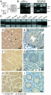
Fig. 1.
Fig. 1.
Generation of mice with conditional knockout of AR in Sertoli cells (S-AR-/y). (A) Mating strategy to generate S-AR-/y mice. (B) Identification and confirmation of S-AR-/y mice. Genomic DNA was isolated from tail snips and used as template for PCR with primers “select and 2–3” and “select and 2–9.” The detailed method and primer sequences have been described (17, 18). The expression of floxed AR, Cre, and internal control IL-2 in the tail genomic DNA of S-AR-/y mice was confirmed by PCR. (C) Genotyping of various tissues harvested from S-AR-/y mice. Only the genomic DNA from the testes of the S-AR-/y mice show the recombinant allele when primers “select and 2–9” are used. (DI) Immunostaining of AR protein in testicular sections from AR+/y, S-AR-/y, and AR-/y mice. (D and E) In AR+/y testis, AR staining was found in Sertoli cells and Leydig cells. (F and G) The S-AR-/y testis shows no AR staining in Sertoli cells; however, Leydig cells retain positive staining (arrows). (H and I) There is no AR staining found in the testis of AR-/y mice. Arrows indicate the location and negative AR staining of Leydig cells. (E, G, and I) The tissue immunostained for AR is counterstained with hematoxylin to reveal the location of cell nuclei.
Fig. 2.
Fig. 2.
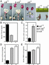
Morphology of testes from 12-wk-old B6 AR+/y, S-AR-/y, and AR-/y mice. (A) Decreased testes size in S-AR-/y mice. S-AR-/y mice have genitourinary organs of similar size as AR+/y mice except for smaller testes, which were one-third the size of AR+/y testes. In contrast, there are small testes but no seminal vesicle, epididymi, or prostate in AR-/y mice. Arrows indicate testes. (B) Serum testosterone levels in 12-wk-old male B6 AR+/y, S-AR-/y, and AR-/y mice. S-AR-/y and AR-/y mice have lower serum testosterone levels than B6 AR+/y mice. (C) Increased expression of ABP is found in S-AR-/y mice. Total RNA was extracted from testes of 12-wk-old AR+/y or S-AR-/y mice and quantitated by real-time PCR (18, 24). The primers for ABP are 5′-CTGCTTCTGCTGTTGCTACTA-3′ and 5′-TTGGTGAGGTCAATGGTCATAAC-3′. Our results indicate increased expression of ABP mRNA in S-AR-/y testis. (D) Compensatory elevation of LH in S-AR-/y mice reveals Leydig cell dysfunction. (E) There was only a marginal change in FSH level in male S-AR-/y and AR-/y mice compared to the age-matched AR+/y mice.
Fig. 3.
Fig. 3.
(A) Analyses of germ cell DNA content of AR+/y and S-AR-/y mice by using flow cytometry. M1 represents haploid cells, M2 represents diploid cells, and M3 represents tetraploid cells. (B) Capα3 expression is absent in S-AR-/y testes. Total RNA from testes of 12-wk-old AR+/y and S-AR-/y mice and quantitated by real-time PCR with Capα3 forward primer 5′-TGGCTCTCAGTTACGCAAGG-3′ and reverse primer 5′-CCAACTCTATCCAAGCACTTACTC-3′.(CH) The maturation of spermatocyte ceases in diplotene stage in S-AR-/y testes. (C) WT testis at tubule stage XI. White arrows indicate diplotene spermatocytes. (D) S-AR-/y testes. No lumen formation is observed in tubules. Some segments of tubule contain many pachytene spermatocytes (white arrowheads). Diplotene spermatocytes (white arrows) can be found in the central region of some tubules. (E) AR-/y testes. Only a small number of pachytene spermatocytes (white arrowheads) can be occasionally seen in the tubules. (F) S-AR-/y testes. Many apoptotic bodies (or degenerate germ cells; black arrowheads) were located in the stage with late pachytene or diplotene spermatocytes. (G) S-AR-/y testes. Meiosis with spindle formation (black arrow) can only rarely be found in the tubules. (H) In S-AR-/y testes, residual secondary spermatocyte (black arrow) and round spermatids (black arrowheads), which have escaped degeneration, are occasionally found in the central region of the tubule.
Fig. 4.
Fig. 4.
Apoptosis and proliferative activity of AR+/y and S-AR-/y testes. (A and B) Proliferative activity of germ cells detected in 12-wk-old male AR+/y and S-AR-/y testes. (C) Quantitation of proliferative cells per seminiferous tubule based on BrdUrd incorporation. (D and E) Terminal deoxynucleotidyltransferase-mediated dUTP nick end labeling (TUNEL) assay to detect the apoptotic cells in 12-wk-old male AR+/y and S-AR-/y testes. (F) Quantitation of apoptotic cells per seminiferous tubule in AR+/y and S-AR-/y testes.
Fig. 5.
Fig. 5.
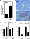
Expression of AMH, cyclin A1, sperm-1, and IGF-1/IGF-1R in AR+/y and S-AR-/y mice. (A) Increased AMH expression in S-AR-/y testes by real-time PCR quantitation. (B) Increased AMH staining in S-AR-/y testis. (C) The expression of IGF-1 and IGF-1R show no significant change in the testes of S-AR-/y mice as compared to age-matched B6 AR+/y mice. (D) Reduced sperm-1 and cyclin A1 expression in S-AR-/y testis by real-time RT-PCR. The detailed real-time PCR method and primer sequences have been described (18). Total RNA and tissue sections were obtained from the testes of 12-wk-old AR+/y and S-AR-/y mice (n = 5 for each group).
Fig. 6.
Fig. 6.
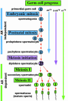
Diagram of germ cell progression in AR+/y,AR-/y, and S-AR-/y testes. AR+/y testes can achieve full germ cell progression. However, spermatogenesis in the AR-/y and S-AR-/y testes ceases predominately in the pachytene and diplotene stages, respectively.

References

    1. Mackay, S. (2000) Int. Rev. Cytol. 200, 47-99. - PubMed
    1. Wolgemuth, D. J., Laurion, E. & Lele, K. M. (2002) Recent Prog. Horm. Res. 57, 75-101. - PubMed
    1. Jegou, B. (1993) Int. Rev. Cytol. 147, 25-96. - PubMed
    1. Roberts, K. P., Awoniyi, C. A., Santulli, R. & Zirkin, B. R. (1991) Endocrinology 129, 3417-3423. - PubMed
    1. Rivarola, M. A., Sanchez, P. & Saez, J. M. (1985) Endocrinology 117, 1796-1802. - PubMed

Publication types