

Dynamic populations of dendritic cell-specific ICAM-3 grabbing nonintegrin-positive immature dendritic cells and liver/lymph node-specific ICAM-3 grabbing nonintegrin-positive endothelial cells in the outer zones of the paracortex of human lymph nodes
- PMID: 15111305
- PMCID: PMC1615649
- DOI: 10.1016/S0002-9440(10)63717-0
Dynamic populations of dendritic cell-specific ICAM-3 grabbing nonintegrin-positive immature dendritic cells and liver/lymph node-specific ICAM-3 grabbing nonintegrin-positive endothelial cells in the outer zones of the paracortex of human lymph nodes
Abstract
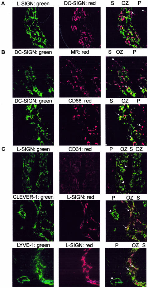
In the paracortex of lymph nodes, cellular immune responses are generated against antigens captured in peripheral tissues by dendritic cells (DCs). DC-SIGN (dendritic cell-specific ICAM-3 grabbing nonintegrin), a C-type lectin exclusively expressed by DCs, functions as an antigen receptor as well as an adhesion receptor. A functional homologue of DC-SIGN, L-SIGN (liver/lymph node-SIGN, also called DC-SIGN-related), is expressed by liver sinus endothelial cells. In lymph nodes, both DC-SIGN and L-SIGN are expressed. In this study, we analyzed the distribution of these two SIGN molecules in detail in both normal and immunoreactive lymph nodes. DC-SIGN is expressed by mature DCs in paracortical areas and in addition by DCs with an immature phenotype in the outer zones of the paracortex. L-SIGN expression was also detected in the outer zones on sinus endothelial cells characterized by their expression of the lymphatic endothelial markers LYVE-1 and CLEVER-1. During both cellular and humoral immune responses changes in the amount of DC-SIGN+ immature and mature DCs and L-SIGN+ endothelial cells were observed, indicating that the influx or proliferation of these cells is dynamically regulated.
Figures


Similar articles
-
Migration of immature mouse DC across resting endothelium is mediated by ICAM-2 but independent of beta2-integrins and murine DC-SIGN homologues.Eur J Immunol. 2006 Oct;36(10):2781-94. doi: 10.1002/eji.200526311. Eur J Immunol. 2006. PMID: 16981228
-
Measles virus targets DC-SIGN to enhance dendritic cell infection.J Virol. 2006 Apr;80(7):3477-86. doi: 10.1128/JVI.80.7.3477-3486.2006. J Virol. 2006. PMID: 16537615 Free PMC article.
-
Dendritic cells recognize tumor-specific glycosylation of carcinoembryonic antigen on colorectal cancer cells through dendritic cell-specific intercellular adhesion molecule-3-grabbing nonintegrin.Cancer Res. 2005 Jul 1;65(13):5935-44. doi: 10.1158/0008-5472.CAN-04-4140. Cancer Res. 2005. PMID: 15994972
-
DC-SIGN (dendritic cell-specific ICAM-grabbing non-integrin) and DC-SIGN-related (DC-SIGNR): friend or foe?Clin Sci (Lond). 2003 Apr;104(4):437-46. Clin Sci (Lond). 2003. PMID: 12653690 Review.
-
DC-SIGN, DC-SIGNR and LSECtin: C-type lectins for infection.Int Rev Immunol. 2014 Jan;33(1):54-66. doi: 10.3109/08830185.2013.834897. Epub 2013 Oct 24. Int Rev Immunol. 2014. PMID: 24156700 Review.
Cited by
-
Role of ChemR23 in directing the migration of myeloid and plasmacytoid dendritic cells to lymphoid organs and inflamed skin.J Exp Med. 2005 Feb 21;201(4):509-15. doi: 10.1084/jem.20041310. J Exp Med. 2005. PMID: 15728234 Free PMC article.
-
Toll-Like Receptor 4 Triggering Promotes Cytosolic Routing of DC-SIGN-Targeted Antigens for Presentation on MHC Class I.Front Immunol. 2018 Jun 14;9:1231. doi: 10.3389/fimmu.2018.01231. eCollection 2018. Front Immunol. 2018. PMID: 29963041 Free PMC article.
-
Role of dendritic cells in the pathogenesis of Whipple's disease.Infect Immun. 2015 Feb;83(2):482-91. doi: 10.1128/IAI.02463-14. Epub 2014 Nov 10. Infect Immun. 2015. PMID: 25385798 Free PMC article.
-
MCP-1 Reduction by L-SIGN Expression in Dengue Virus-Infected Liver Endothelial Cells.Viruses. 2025 Feb 28;17(3):344. doi: 10.3390/v17030344. Viruses. 2025. PMID: 40143272 Free PMC article.
-
Recent advances in DENV receptors.ScientificWorldJournal. 2013 Apr 23;2013:684690. doi: 10.1155/2013/684690. Print 2013. ScientificWorldJournal. 2013. Retraction in: ScientificWorldJournal. 2013 Nov 18;2013:235198. doi: 10.1155/2013/235198. PMID: 23737723 Free PMC article. Retracted. Review.
References
-
- Banchereau J, Steinman RM. Dendritic cells and the control of immunity. Nature. 1998;392:245–252. - PubMed
-
- Lanzavecchia A, Sallusto F. Regulation of T cell immunity by dendritic cells. Cell. 2001;106:263–266. - PubMed
-
- Gretz JE, Norbury CC, Anderson AO, Proudfoot AE, Shaw S. Lymph-borne chemokines and other low molecular weight molecules reach high endothelial venules via specialized conduits while a functional barrier limits access to the lymphocyte microenvironments in lymph node cortex. J Exp Med. 2000;192:1425–1440. - PMC - PubMed
-
- Salomon B, Cohen JL, Masurier C, Klatzmann D. Three populations of mouse lymph node dendritic cells with different origin and dynamics. J Immunol. 1998;160:708–717. - PubMed
Publication types
MeSH terms
Substances
LinkOut - more resources
Full Text Sources
Miscellaneous
