

Nonequilibrium behavior in supported lipid membranes containing cholesterol
- PMID: 15111410
- PMCID: PMC1304162
- DOI: 10.1016/S0006-3495(04)74345-3
Nonequilibrium behavior in supported lipid membranes containing cholesterol
Abstract
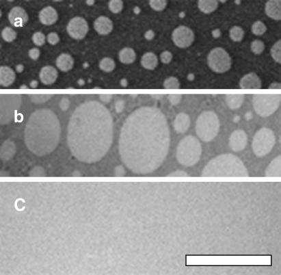
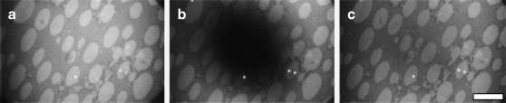
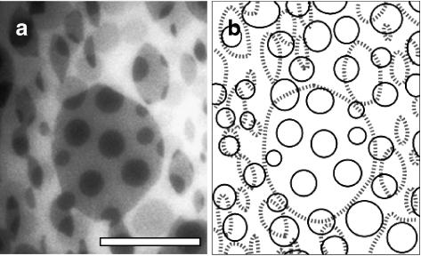
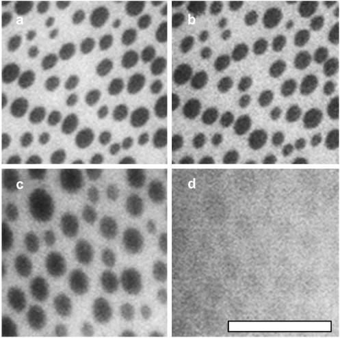
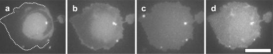
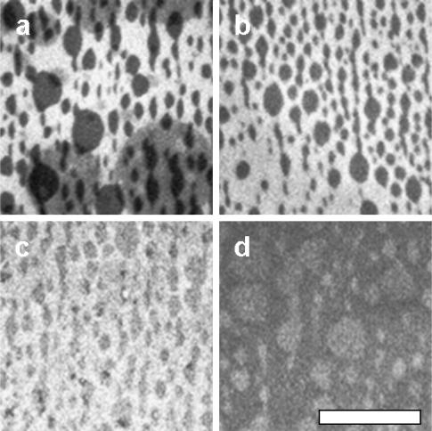
We investigate lateral organization of lipid domains in vesicles versus supported membranes and monolayers. The lipid mixtures used are predominantly DOPC/DPPC/Chol and DOPC/BSM/Chol, which have been previously shown to produce coexisting liquid phases in vesicles and monolayers. In a monolayer at an air-water interface, these lipids have miscibility transition pressures of approximately 12-15 mN/m, which can rise to 32 mN/m if the monolayer is exposed to air. Lipid monolayers can be transferred by Langmuir-Schäfer deposition onto either silanized glass or existing Langmuir-Blodgett supported monolayers. Micron-scale domains are present in the transferred lipids only if they were present in the original monolayer before deposition. This result is valid for transfers at 32 mN/m and also at lower pressures. Domains transferred to glass supports differ from liquid domains in vesicles because they are static, do not align in registration across leaflets, and do not reappear after temperature is cycled. Similar static domains are found for vesicles ruptured onto glass surfaces. Although supported membranes on glass capture some aspects of vesicles in equilibrium (e.g., gel-liquid transition temperatures and diffusion rates of individual lipids), the collective behavior of lipids in large liquid domains is poorly reproduced.
Figures

Similar articles
-
Miscibility of ternary mixtures of phospholipids and cholesterol in monolayers, and application to bilayer systems.Biophys J. 2005 Jan;88(1):269-76. doi: 10.1529/biophysj.104.048439. Epub 2004 Oct 8. Biophys J. 2005. PMID: 15475588 Free PMC article.
-
Liquid-liquid phase transition temperatures increase when lipid bilayers are supported on glass.Biochim Biophys Acta Biomembr. 2018 Oct;1860(10):1965-1971. doi: 10.1016/j.bbamem.2018.05.001. Epub 2018 May 10. Biochim Biophys Acta Biomembr. 2018. PMID: 29752899
-
Organization in lipid membranes containing cholesterol.Phys Rev Lett. 2002 Dec 23;89(26):268101. doi: 10.1103/PhysRevLett.89.268101. Epub 2002 Dec 9. Phys Rev Lett. 2002. PMID: 12484857
-
Properties of Langmuir and solid supported lipid films with sphingomyelin.Adv Colloid Interface Sci. 2015 Aug;222:385-97. doi: 10.1016/j.cis.2014.03.008. Epub 2014 Mar 28. Adv Colloid Interface Sci. 2015. PMID: 24725646 Review.
-
Seeing spots: complex phase behavior in simple membranes.Biochim Biophys Acta. 2005 Dec 30;1746(3):172-85. doi: 10.1016/j.bbamcr.2005.06.010. Epub 2005 Jul 6. Biochim Biophys Acta. 2005. PMID: 16043244 Review.
Cited by
-
Dilatational and shear rheology of soluble and insoluble monolayers with a Langmuir trough.J Colloid Interface Sci. 2023 Jan;629(Pt A):125-135. doi: 10.1016/j.jcis.2022.08.051. Epub 2022 Aug 13. J Colloid Interface Sci. 2023. PMID: 36063630 Free PMC article.
-
Characterization of the ordered phase formed by sphingomyelin analogues and cholesterol binary mixtures.Biophysics (Nagoya-shi). 2013 May 22;9:37-49. doi: 10.2142/biophysics.9.37. eCollection 2013. Biophysics (Nagoya-shi). 2013. PMID: 27493539 Free PMC article.
-
Main phase transitions in supported lipid single-bilayer.Biophys J. 2005 Aug;89(2):1094-101. doi: 10.1529/biophysj.105.062463. Epub 2005 May 6. Biophys J. 2005. PMID: 15879467 Free PMC article.
-
Miscibility of ternary mixtures of phospholipids and cholesterol in monolayers, and application to bilayer systems.Biophys J. 2005 Jan;88(1):269-76. doi: 10.1529/biophysj.104.048439. Epub 2004 Oct 8. Biophys J. 2005. PMID: 15475588 Free PMC article.
-
Galactosylceramide domain microstructure: impact of cholesterol and nucleation/growth conditions.Biophys J. 2006 Jun 15;90(12):4466-78. doi: 10.1529/biophysj.105.072744. Epub 2006 Mar 24. Biophys J. 2006. PMID: 16565044 Free PMC article.
References
-
- Anderson, R. G., and K. Jacobson. 2002. A role for lipid shells in targeting proteins to caveolae, rafts, and other lipid domains. Science. 296:1821–1825. - PubMed
-
- Benvegnu, D. J., and H. M. McConnell. 1993. Surface dipole densities in lipid monolayers. J. Phys. Chem. 97:6686–6691.
-
- Bezzine, S., J. G. Bollinger, A. G. Singer, S. L. Veatch, S. L. Keller, and M. H. Gelb. 2002. On the binding preference of human groups IIA and X phospholipases A2 for membranes with anionic phospholipids. J. Biol. Chem. 277:48523–48534. - PubMed
-
- Cornell, B. A., V. L. Braach-Maksvytis, L. G. King, P. D. Osman, B. Raguse, L. Wieczorek and R. J. Pace. 1997. A biosensor that uses ion-channel switches. Nature. 387:580–583. - PubMed
-
- de Jong, K., D. Geldwerth, and F. A. Kuypers. 1997. Oxidative damage does not alter membrane phospholipid asymmetry in human erythrocytes. Biochemistry. 36:6768–6776. - PubMed
Publication types
MeSH terms
Substances
Grants and funding
LinkOut - more resources
Full Text Sources
Medical
