. 2004 Mar 16:5:5.
doi: 10.1186/1471-2172-5-5.

Endothelial cells present antigens in vivo

Affiliations

Endothelial cells present antigens in vivo

Annette L Rothermel et al. BMC Immunol. .

Abstract

Background: Immune recognition of vascular endothelial cells (EC) has been implicated in allograft rejection, protection against pathogens, and lymphocyte recruitment. However, EC pervade nearly all tissues and predominate in none, complicating any direct test of immune recognition. Here, we examined antigen presentation by EC in vivo by testing immune responses against E. coli beta-galactosidase (beta-gal) in two lines of transgenic mice that express beta-gal exclusively in their EC. TIE2-lacZ mice express beta-gal in all EC and VWF-lacZ mice express beta-gal in heart and brain microvascular EC.

Results: Transgenic and congenic wild type FVB mice immunized with beta-gal expression vector DNA or beta-gal protein generated high titer, high affinity antisera containing comparable levels of antigen-specific IgG1 and IgG2a isotypes, suggesting equivalent activation of T helper cell subsets. The immunized transgenic mice remained healthy, their EC continued to express beta-gal, and their blood vessels showed no histological abnormalities. In response to beta-gal in vitro, CD4+ and CD8+ T cells from immunized transgenic and FVB mice proliferated, expressed CD25, and secreted IFN-gamma. Infection with recombinant vaccinia virus encoding beta-gal raised equivalent responses in transgenic and FVB mice. Hearts transplanted from transgenic mice into FVB mice continued to beat and the graft EC continued to express beta-gal. These results suggested immunological ignorance of the transgene encoded EC protein. However, skin transplanted from TIE2-lacZ onto FVB mice lost beta-gal+ EC and the hosts developed beta-gal-specific antisera, demonstrating activation of host immune effector mechanisms. In contrast, skin grafted from TIE2-lacZ onto VWF-lacZ mice retained beta-gal+ EC and no antisera developed, suggesting a tolerant host immune system.

Conclusion: Resting, beta-gal+ EC in transgenic mice tolerize specific lymphocytes that would otherwise respond against beta-gal expressed by EC within transplanted skin. We conclude that EC effectively present intracellular "self" proteins to the immune system. However, antigen presentation by EC does not delete or anergize a large population of specific lymphocytes that respond to the same protein following conventional immunization with protein or expression vector DNA. These results clearly demonstrate striking context sensitivity in the immune recognition of EC, a subtlety that must be better understood in order to treat immune diseases and complications involving the vasculature.


Figures

Figure 1
Figure 1
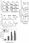
Genetic immunization with a β-gal expression vector induces strong humoral responses in wild type mice (FVB) and transgenic mice (VWF-lacZ and TIE2-lacZ) that express β-gal in their endothelial cells A. Equivalent β-gal specific antisera are generated in FVB and VWF-lacZ mice. ELISAs were performed on antisera from mice immunized 6 weeks earlier with a gene gun. Closed markers represent sera from mice immunized with the expression vector CMV-β gal. Open markers represent preimmune sera (VWF-lacZ) or sera from mice immunized with a control expression vector (CMV-GFP, FVB). Similar results were obtained in two independent experiments. B. The humoral response matures in wild type and transgenic mice. Titers of antibodies in control sera (0) and sera from mice immunized once (1') or twice (2') with the CMV-β-gal expression vector DNA are shown. Similar results were obtained in two independent experiments. C. TIE2-lacZ mice respond against β-gal. Antisera of wild type (FVB) and TIE2-lacZ mice immunized once 6 weeks previously are shown. Closed symbols represent sera assayed on plates coated with β-gal, open symbols represent assays on control, BSA-coated plates. Similar results were obtained in two independent experiments. Titers of sera from pre- or un-immunized mice were <10-2 (not shown). D. Soluble β-gal competes for antibodies in sera from FVB and transgenic mice equivalently. Sera from immunized and boosted mice were mixed with soluble β-gal at the concentrations indicated and incubated overnight at 4°C. Free antibodies were then detected by adding the mixture to β-gal coated wells.
Figure 2
Figure 2
Immunization with β-gal protein in adjuvant raises strong antibody responses in wild type (FVB) and transgenic (VWF-lacZ and TIE2-lacZ) mice. Serum samples were taken 2 weeks after boosting with soluble β-gal and specific antibodies were measured by ELISA. Closed symbols represent sera assayed on β-gal coated plates and open symbols represent sera assayed on control, BSA-coated plates.
Figure 3
Figure 3
T cells from immunized FVB and transgenic VWF-lacZ and TIE2-lacZ mice respond to β-gal in vitro. A. CD4+ and CD8+ lymph node T cells proliferate and express IL-2 receptors (CD25+) in response to β-gal. Representative FACS profiles are shown. Lymph node cells were obtained from mice 7 days after in vivo priming in all the experiments shown. Lymph node cells (2 × 10⁵) were cultured in flat bottom wells and stained for CD4, CD8, and CD25 on day 4. The histograms represent CD25 expression in cultures receiving β-gal (thick line) or not (thin line). B. CD25+ lymphocyte populations expand in cultures of lymph node cells from FVB and transgenic mice. The results of three independent FACS experiments are shown, each represented with a different symbol, and the mean response at each time is indicated with a dash. For the stimulation indices, the background proliferation of cultures receiving no addition or ovalbumin additions were averaged. C. Wild type and transgenic T cells display equivalent avidity for β-gal. Lymph node cells (105) were cultured with irradiated (1500 R) splenocytes (106) in flat bottom wells with the indicated concentrations of β-gal. Results for day 4 are shown. Similar results were obtained in more than 4 independent dose-response proliferation assays. D. FVB and transgenic T cells secrete IFN-γ in response to β-gal. Lymph node cells (2 × 10⁵) were cultured in round bottom wells with β-gal (closed squares) or without (open squares). Culture superantants were recovered at the times indicated, one sample per culture, three cultures per point, and IFN-γ was measured by ELISA.
Figure 4
Figure 4
EC continue to express β-gal in transgenic VWF-lacZ and TIE2-lacZ mice mounting strong immune responses against β-gal. A. EC in cardiac vessels of immunized, transgenic mice continue to make β-gal. Control, unimmunized mice and mice immunized and boosted with β-gal protein were sacrificed and their hearts were harvested and stained with X-gal. Representative fields are shown. Similar results were obtained in four independent experiments, two analyzing mice immunized with β-gal protein and two analyzing mice immunized with β-gal expression vectors. B. Dermal EC of immunized, transgenic TIE2-lacZ mice continue to express β-gal. Mice were sacrificed and full-thickness portions of their skins were stained with X-gal. The underside is shown. Unimmunized TIE2-lacZ mice expressed equivalent levels of β-gal, as assessed by X-gal staining (not shown).
Figure 5
Figure 5
Transplanted hearts from transgenic mice are tolerated and continue to express β-gal. Heterotopic transplants were harvested 3 months after transplantation and stained with X-gal. Similar results were obtained in 2 additional experiments.
Figure 6
Figure 6
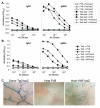
β-gal+ EC trigger immune responses and are cleared from TIE2-lacZ skin grafts onto wild type FVB but not VWF-lacZ hosts. A. ELISA of sera from reciprocal donor-host skin transplants detects β-gal specific antibodies in FVB but not TIE2-lacZ hosts. Similar results were obtained in 3 separate experiments, in which 7 out of 8 wild type mice generated strong IgG2a isotype antibodies to β-gal following TIE2-lacZ skin grafts. B. VWF-lacZ mice do not raise β-gal-specific antisera following grafting with TIE2-lacZ skin. Grafting and antisera were analyzed as described above.C. EC expressing β-gal are cleared from TIE2-lacZ skin grafts onto host wild type FVB but not VWF-lacZ mice. Photomicrographs of the underside of X-gal stained donor TIE2-lacZ skin and TIE2-lacZ skin grafts from FVB or VWF-lacZ hosts are shown.

Similar articles

Cited by

References

    1. Pober JS, Cotran RS. The role of endothelial cells in inflammation. Transplantation. 1990;50:537–544. - PubMed
    1. Austrup F, Vestweber D, Borges E, Löhning M, Bräuer R, Herz U, Renz H, Hallmann R, Scheffold A, Radbruch A, Hamann A. P- and E-selectin mediate recruitment of T-helper-1 but not T-helper-2 cells into inflamed tissues. Nature. 1997;385:81–83. doi: 10.1038/385081a0. - DOI - PubMed
    1. de Waal RM, Bogman MJ, Maass CN, Cornelissen LM, Tax WJ, Koene RA. Variable expression of Ia antigens on the vascular endothelium of mouse skin allografts. Nature. 1983;303:426–429. - PubMed
    1. Racusen LC, Solez K, Colvin RB, Bonsib SM, Castro MC, Cavallo T, Croker BP, Demetris AJ, Drachenberg CB, Fogo AB, Furness P, Gaber LW, Gibson IW, Glotz D, Goldberg JC, Grande J, Halloran PF, Hansen HE, Hartley B, Hayry PJ, Hill CM, Hoffman EO, Hunsicker LG, Lindblad AS, Yamaguchi Y, et al. The Banff 97 working classification of renal allograft pathology. Kidney Int. 1999;55:713–723. doi: 10.1046/j.1523-1755.1999.00299.x. - DOI - PubMed
    1. Ross R. Atherosclerosis – an inflammatory disease. N Engl J Med. 1999;340:115–126. doi: 10.1056/NEJM199901143400207. - DOI - PubMed

Publication types

MeSH terms